. 2002 Jul 8;158(1):23-9.
doi: 10.1083/jcb.200202053. Epub 2002 Jul 8.

Rapid exchange of mammalian topoisomerase II alpha at kinetochores and chromosome arms in mitosis

Affiliations

Rapid exchange of mammalian topoisomerase II alpha at kinetochores and chromosome arms in mitosis

Penny A Tavormina et al. J Cell Biol. .

Abstract

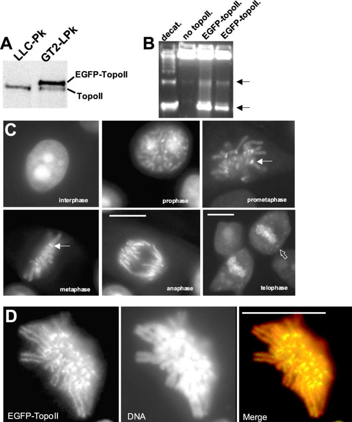
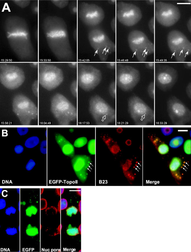
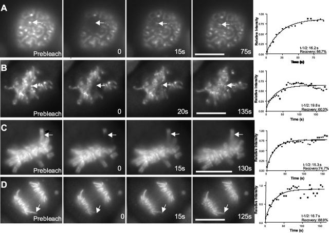
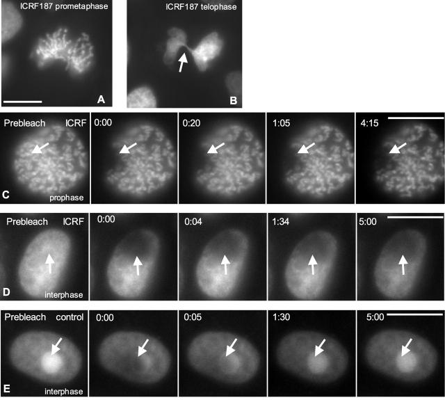
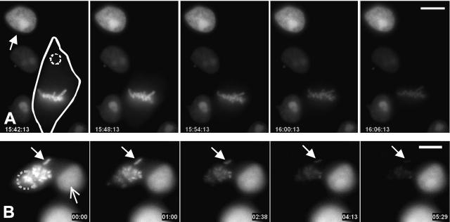
A stable cell line (GT2-LPk) derived from LLC-Pk was created in which endogenous DNA topoisomerase II alpha (topoII alpha) protein was downregulated and replaced by the expression of topoII alpha fused with enhanced green fluorescent protein (EGFP-topoII alpha). The EGFP-topoII alpha faithfully mimicked the distribution of the endogenous protein in both interphase and mitosis. In early stages of mitosis, EGFP-topoII alpha accumulated at kinetochores and in axial lines extending along the chromosome arms. During anaphase, EGFP-topoII alpha diminished at kinetochores and increased in the cytoplasm with a portion accumulating into large circular foci that were mobile and appeared to fuse with the reforming nuclei. These cytoplasmic foci appearing at anaphase were coincident with precursor organelles of the reforming nucleolus called nucleolus-derived foci (NDF). Photobleaching of EGFP-topoII alpha associated with kinetochores and chromosome arms showed that the majority of the protein rapidly exchanges (t1/2 of 16 s). Catalytic activity of topoII alpha was essential for rapid dynamics, as ICRF-187, an inhibitor of topoII alpha, blocked recovery after photobleaching. Although some topoII alpha may be stably associated with chromosomes, these studies indicate that the majority undergoes rapid dynamic exchange. Rapid mobility of topoII alpha in chromosomes may be essential to resolve strain imparted during chromosome condensation and segregation.


Figures

Figure 1.
Figure 1.
GT2-LPk cells express EGFP-topoIIα. (A) Immunoblotting with anti-topoIIα antibody of whole-cell extracts of parental LLC-Pk cells and GT2-LPk. GT2-LPk cells show downregulated expression of endogenous topoIIα. (B) EGFP–topoIIα immunoprecipitated from GT2-LPk cells catalyzes decatenation of kinetoplast DNA. Lanes contain respectively decatenated control, kinetoplast DNA with no topoisomerase II added, and two separate immunoprecipitations of GT2-LPk extracts with anti-GFP antibody. Arrows indicate the migration of the decatenated products (nicked and supercoiled). (C) EGFP– topoIIα localization in a panel of living cells at various stages of mitosis. EGFP–topoIIα associates with chromosomes in prophase, becomes concentrated at kinetochores (closed arrows) and along chromosome arms in prometaphase and metaphase, and associates with chromosome arms in anaphase. In late anaphase and telophase, EGFP–topoIIα increases in the cytoplasm with some becoming concentrated in cytoplasmic foci (open arrow). (D) Imaging of fixed GT2-LPk cell at late prometaphase after staining with DAPI shows localization of topoisomerase II to the kinetochore region and axes of chromosomes. Cells were simultaneously fixed and permeabilized by treatment with 2% formaldehyde, 0.5% TX-100 in microtubule stabilizing buffer (60 mM Pipes, 25 mM Hepes, 10 mM EGTA, 4 mM MgSO4, pH 6.95). The images represent a maximum projection of six 0.2-μm optical sections. In the merged image, EGFP–topoIIα is green and appears yellow due to overlap with the DNA in red. Bars, 10 μm.
Figure 2.
Figure 2.
EGFP–topoIIα localization is dynamic at anaphase and telophase. (A) Selected frames from a time-lapse video of a GT2-LPk cell in late mitosis. In anaphase, some of the cytoplasmic EGFP–topoIIα concentrates into distinct foci (closed arrows) that are motile. At telophase, the foci appear to fuse with the reforming nucleus (open arrows), whereas nucleoli become more apparent. (Image frames in this sequence were individually adjusted for contrast to optimize visualization of the cyto- plasmic foci; Video 1, available at http://www.jcb.org/cgi/content/full/jcb.200202053/DC1) (B) EGFP–topoIIα foci (arrows) in telophase cells colocalize with NDFs identified by labeling with antibody to nucleolar B23 protein, a gift from Dr. Mirek Dundr (National Cancer Institute, Bethesda, MD). (The EGFP– topoIIα image is oversaturated for the nuclear fluorescence to clearly visualize the cytoplasmic foci.) (C) EGFP–topoIIα foci do not colocalize with reforming nuclear envelope identified by RL1 antibody to nuclear pore proteins, a gift from Dr. Bryce Pascal (University of Virginia, Charlottesville, VA). Bars, 10 μm.
Figure 3.
Figure 3.
Recovery of EGFP–topoIIα fluorescence after photobleaching occurs rapidly at kinetochores and chromosome arms. Examples showing targeting of kinetochores (arrows) at prophase (A) and prometaphase (B) and targeting of chromosome arms (arrows) at metaphase (C) and anaphase (D) Images include a prebleach image, one taken just after photobleaching (0 time) and two images taken during recovery. Graphs at the end of each sequence show fitting of corrected intensity measurements in the photobleached regions to determine recovery half times (t-1/2) and total degree of recovery (recovery). Bars, 10 μm.
Figure 4.
Figure 4.
Catalytic activity of topoIIα is required for rapid recovery after photobleaching. (A and B) GT2-LPk cells treated with ICRF-187, an inhibitor of topoIIα, showed impaired chromosome condensation in prometaphase (A) and mitotic catastrophe as the cleavage furrow has cut through the mass of unseparated chromatids in telophase (B, arrow). (C) Photobleaching of mitotic cells shows very limited recovery (arrow). (D) Photobleaching of interphase nucleus of ICRF-187–treated cell also shows limited recovery. ICRF-187 treatment reduces the concentration of EGFP-topoIIα at the nucleoli. (E) Photobleaching of nondrug treated interphase cell nucleus results in rapid recovery (arrow). Bars, 10 μm.
Figure 5.
Figure 5.
Fluorescence loss in photobleaching demonstrates exchange of topoIIα between chromosomal and cytoplasmic pools in mitotic cells. (A) Exchange of topoIIα occurs between the chromosomes and the cytoplasm of mitotic cells. A late prometaphase cell outlined by the solid line was targeted for photobleaching in the cytoplasm away from the chromosomes indicated (dotted circle). The region was photobleached every 20 s and images were captured every 2 min. Repeated photobleaching of the cytoplasm results in depletion of fluorescence in the chromosomes. The fluorescence intensity of the chromosomes was reduced by 79%, whereas that of the control interphase nucleus (arrow) in the adjacent interphase cell was reduced only 8%. (B) Exchange of topoIIα occurs through the cytoplasm among chromosomes. A cell treated with 0.1 μg/ml nocodazole was repeatedly bleached in the lower region of the mass of chromosomes (dotted circle). Fluorescence intensity of the isolated chromosome at top (closed arrows) diminished by 85%, whereas the intensity of the nucleus in the neighboring cell (open arrow) decreased by only 5% over the course of the observations (Video 2, available at http://www.jcb.org/cgi/content/full/jcb.200202053/DC1). Bars, 10 μm.

References

    1. Bakshi, R.P., S. Galande, and K. Muniyappa. 2001. Functional and regulatory characteristics of eukaryotic type II DNA topoisomerase. Crit. Rev. Biochem. Mol. Biol. 36:1–37. - PubMed
    1. Barthelmes, H.U., P. Grue, S. Feineis, T. Straub, and F. Boege. 2000. Active DNA topoisomerase IIα is a component of the salt-stable centrosome core. J. Biol. Chem. 275:38823–38830. - PubMed
    1. Christensen, M.O., M.K. Larsen, H.U. Barthelmes, R. Hock, C.L. Andersen, E. Kjeldsen, B.R. Knudsen, O. Westergaard, F. Boege, and C. Mielke. 2002. Dynamics of human DNA topoisomerases IIα and IIβ in living cells. J. Cell Biol. 157:31–44. - PMC - PubMed
    1. Dundr, M., T. Misteli, and M.O. Olson. 2000. The dynamics of postmitotic reassembly of the nucleolus. J. Cell Biol. 150:433–446. - PMC - PubMed
    1. Earnshaw, W.C., B. Halligan, C.A. Cooke, M.M. Heck, and L.F. Liu. 1985. Topoisomerase II is a structural component of mitotic chromosome scaffolds. J. Cell Biol. 100:1706–1715. - PMC - PubMed

Publication types

MeSH terms