

Dual-wavelength fluorescent speckle microscopy reveals coupling of microtubule and actin movements in migrating cells
- PMID: 12105180
- PMCID: PMC2173033
- DOI: 10.1083/jcb.200203022
Dual-wavelength fluorescent speckle microscopy reveals coupling of microtubule and actin movements in migrating cells
Abstract
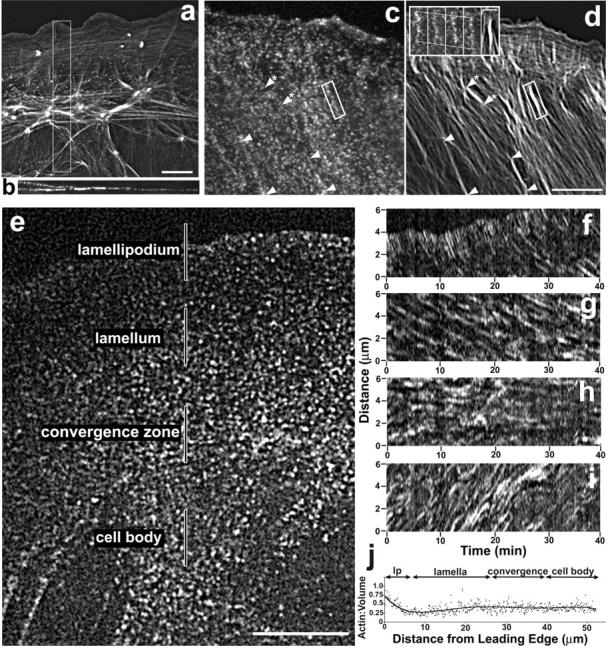
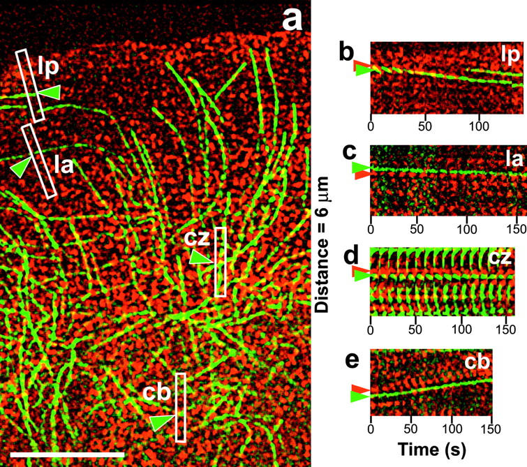
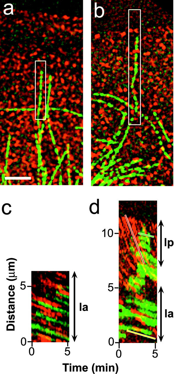
Interactions between microtubules (MTs) and filamentous actin (f-actin) are involved in directed cell locomotion, but are poorly understood. To test the hypothesis that MTs and f-actin associate with one another and affect each other's organization and dynamics, we performed time-lapse dual-wavelength spinning-disk confocal fluorescent speckle microscopy (FSM) of MTs and f-actin in migrating newt lung epithelial cells. F-actin exhibited four zones of dynamic behavior: rapid retrograde flow in the lamellipodium, slow retrograde flow in the lamellum, anterograde flow in the cell body, and no movement in the convergence zone between the lamellum and cell body. Speckle analysis showed that MTs moved at the same trajectory and velocity as f-actin in the cell body and lamellum, but not in the lamellipodium or convergence zone. MTs grew along f-actin bundles, and quiescent MT ends moved in association with f-actin bundles. These results show that the movement and organization of f-actin has a profound effect on the dynamic organization of MTs in migrating cells, and suggest that MTs and f-actin bind to one another in vivo.
Figures

Similar articles
-
Converging populations of f-actin promote breakage of associated microtubules to spatially regulate microtubule turnover in migrating cells.Curr Biol. 2002 Nov 19;12(22):1891-9. doi: 10.1016/s0960-9822(02)01276-9. Curr Biol. 2002. PMID: 12445381
-
Simultaneous mapping of filamentous actin flow and turnover in migrating cells by quantitative fluorescent speckle microscopy.Proc Natl Acad Sci U S A. 2004 Jun 29;101(26):9660-5. doi: 10.1073/pnas.0300552101. Epub 2004 Jun 21. Proc Natl Acad Sci U S A. 2004. PMID: 15210979 Free PMC article.
-
Actomyosin-based retrograde flow of microtubules in the lamella of migrating epithelial cells influences microtubule dynamic instability and turnover and is associated with microtubule breakage and treadmilling.J Cell Biol. 1997 Oct 20;139(2):417-34. doi: 10.1083/jcb.139.2.417. J Cell Biol. 1997. PMID: 9334345 Free PMC article.
-
Coupled zones of f-actin and microtubule movement in polarized cells.Dev Cell. 2002 Aug;3(2):152-3. doi: 10.1016/s1534-5807(02)00227-7. Dev Cell. 2002. PMID: 12194844 Review.
-
Quantitative fluorescent speckle microscopy: where it came from and where it is going.J Microsc. 2003 Sep;211(Pt 3):191-207. doi: 10.1046/j.1365-2818.2003.01222.x. J Microsc. 2003. PMID: 12950468 Review.
Cited by
-
Asymmetric formation of coated pits on dorsal and ventral surfaces at the leading edges of motile cells and on protrusions of immobile cells.Mol Biol Cell. 2015 Jun 1;26(11):2044-53. doi: 10.1091/mbc.E15-01-0055. Epub 2015 Apr 7. Mol Biol Cell. 2015. PMID: 25851602 Free PMC article.
-
Rho-dependent formation of epithelial "leader" cells during wound healing.Proc Natl Acad Sci U S A. 2003 Sep 16;100(19):10788-93. doi: 10.1073/pnas.1834401100. Epub 2003 Sep 5. Proc Natl Acad Sci U S A. 2003. PMID: 12960404 Free PMC article.
-
Differential RhoA dynamics in migratory and stationary cells measured by FRET and automated image analysis.PLoS One. 2008;3(12):e4082. doi: 10.1371/journal.pone.0004082. Epub 2008 Dec 30. PLoS One. 2008. PMID: 19114999 Free PMC article.
-
A cytoskeletal clutch mediates cellular force transmission in a soft, three-dimensional extracellular matrix.Mol Biol Cell. 2017 Jul 7;28(14):1959-1974. doi: 10.1091/mbc.E17-02-0102. Epub 2017 Jun 7. Mol Biol Cell. 2017. PMID: 28592635 Free PMC article.
-
Finding the cell center by a balance of dynein and myosin pulling and microtubule pushing: a computational study.Mol Biol Cell. 2010 Dec;21(24):4418-27. doi: 10.1091/mbc.E10-07-0627. Epub 2010 Oct 27. Mol Biol Cell. 2010. PMID: 20980619 Free PMC article.
References
-
- Cramer, L.P. 1999. Role of actin-filament disassembly in lamellipodium protrusion in motile cells revealed using the drug jasplakinolide. Curr. Biol. 9:1095–1105. - PubMed
-
- Etienne-Manneville, S., and A. Hall. 2001. Integrin-mediated activation of Cdc42 controls cell polarity in migtating astrocytes through PKCξ. Cell. 106:489–498. - PubMed
-
- Fuchs, E., and I. Karakesisoglou. 2001. Bridging cytoskeletal intersections. Gene Dev. 15:1–14. - PubMed
Publication types
MeSH terms
Substances
Grants and funding
LinkOut - more resources
Full Text Sources
Other Literature Sources
