

The vasculature and its role in the damaged and healing tendon
- PMID: 12106496
- PMCID: PMC128932
- DOI: 10.1186/ar416
The vasculature and its role in the damaged and healing tendon
Abstract
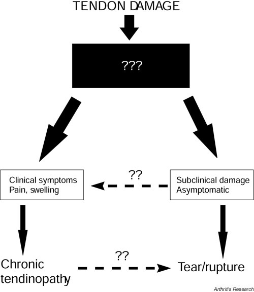
Tendon pathology has many manifestations, from spontaneous rupture to chronic tendinitis or tendinosis; the etiology and pathology of each are very different, and poorly understood. Tendon is a comparatively poorly vascularised tissue that relies heavily upon synovial fluid diffusion to provide nutrition. During tendon injury, as with damage to any tissue, there is a requirement for cell infiltration from the blood system to provide the necessary reparative factors for tissue healing. We describe in this review the response of the vasculature to tendon damage in a number of forms, and how and when the revascularisation or neovascularisation process occurs. We also include a section on the revascularisation of tendon during its use as a tendon graft in both ligament reconstruction and tendon-tendon grafting.
Figures
References
-
- Manske PR, Lesker PA. Biochemical evidence of flexor tendon participation in the repair process – an in vitro study. J Hand Surg. 1984;9B:117–120. - PubMed
-
- Gelberman RH, Manske PR, Van de Berg JS, Lesker PA, Akeson WH. Flexor tendon repair. in-vitro: 1984;a comparitive histologic study of the rabbit, chicken, dog and monkey:39–48. - PubMed
-
- Potenza AD. Tendon healing within the flexor digital sheath in the dog. J Bone Joint Surg. 1962;44A:49–64. - PubMed
-
- Gelberman RH, Manske PR, Akeson WH, Woo SL, Lundborg G, Amiel D. Flexor tendon repair. J Orthop Res. 1986;4:119–128. - PubMed
-
- Li BH, Hong GX, Zu TB. A histological observation on the flexor tendon healing within intact sheath. J Tongji Med Univ. 1991;11:169–173. - PubMed
Publication types
MeSH terms
LinkOut - more resources
Full Text Sources
Other Literature Sources
Medical
