. 2002 Jul;161(1):89-96.
doi: 10.1016/S0002-9440(10)64160-0.

Microinjection of cathepsin d induces caspase-dependent apoptosis in fibroblasts

Affiliations

Microinjection of cathepsin d induces caspase-dependent apoptosis in fibroblasts

Karin Roberg et al. Am J Pathol. 2002 Jul.

Abstract

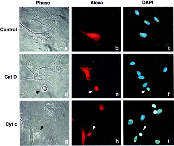
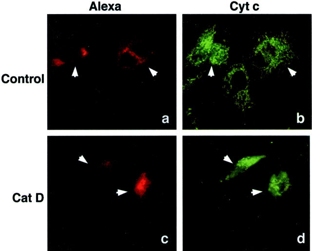
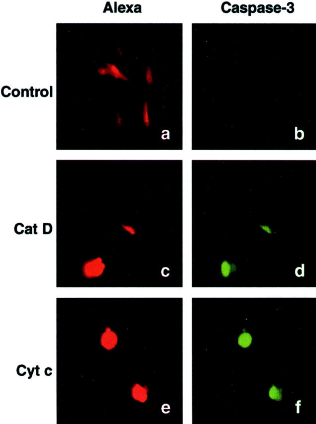
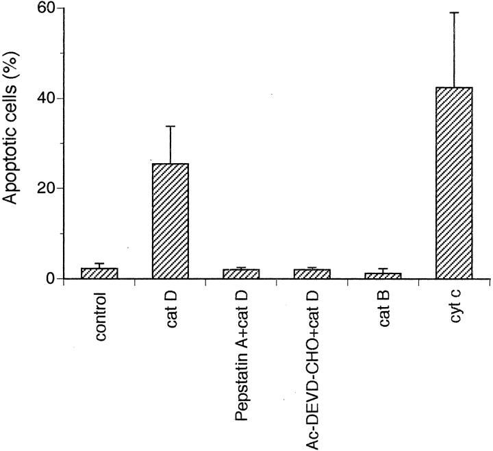
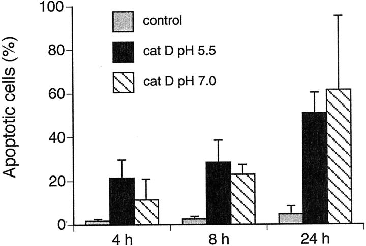
Recent reports have indicated that enzymes such as cathepsins D and B are translocated from lysosomal compartments to the cytosol early during apoptosis. We have previously noted that a translocation of cathepsins D and B occur before cytochrome c release and caspase activation in cardiomyocytes and human fibroblasts during oxidative stress-induced apoptosis. In the present report, we use a microinjection technique to investigate if cytosolic location of the cathepsins D and B are important for induction of apoptosis. We found that microinjection of cathepsin D into the cytosol of human fibroblasts caused apoptosis, which was detected as changes in distribution of cytochrome c, cell shrinkage, activation of caspases, chromatin condensation, and formation of pycnotic nuclei. No apoptosis was, however, induced by microinjection of cathepsin B. Moreover, apoptosis was prevented in fibroblasts pretreated with a caspase-3-like inhibitor, and also when microinjected with cathepsin D mixed with the cathepsin D inhibitor, pepstatin A. These results show that cytosolic cathepsin D can act as a proapoptotic mediator upstream of cytochrome c release and caspase activation in human fibroblasts.


Figures

Figure 1.
Figure 1.
Light and fluorescence microscopy of fibroblasts 8 hours after microinjection of cat D or cyt c. Apoptotic fibroblasts were detected as shrunken cells with pycnotic nuclei by light and fluorescence microscopy. a–c: Cells microinjected with Alexa Fluor alone. No apoptosis was detected. d–f: Cells microinjected with Alexa Fluor together with 0.25 mg/ml of cat D, or g–i, together with 3 mg/ml of cyt c. Microinjected cells were visualized with Alexa Fluor (b, e, and h; red fluorescence) and nuclear morphology by 4,6-diamidino-2-phenylindole staining (c, f, and i). Arrows show microinjected apoptotic cells.
Figure 2.
Figure 2.
Transmission electron microscopy of microinjected fibroblasts. The micrographs show fibroblasts 6 hours after microinjection with Alexa Fluor alone (A) or with Alexa Fluor and 3 mg/ml of cyt c (B), or with Alexa Fluor and 0.25 mg/ml of cat D (C and D). Note in A a normal nucleus (asterisk) and in B–D apoptotic morphology-like partially fragmented nuclei, nuclei with condensed chromatin (arrows), and condensed mitochondria around the nucleus (arrowheads). Original magnifications: ×2000 (A, B, and D); ×1000 (C). Scale bars: 4 μm (A, B, and D); 10 μm (C).
Figure 3.
Figure 3.
Cyt c location by immunofluorescence staining of fibroblasts. Micrographs of fibroblasts 2 hours after microinjection with Alexa Fluor alone (a and b) or with Alexa Fluor together with 0.25 mg/ml of cat D (c and d). Microinjected cells detected by the red fluorescence of Alexa Fluor (a and c), and cyt c by immunofluorescence staining with a FITC-conjugated secondary antibody (b and d). Arrows indicate microinjected cells.
Figure 4.
Figure 4.
Caspase-3-like protease activity was detected 4 hours after microinjection, using a FITC conjugate of the caspase inhibitor VAD-FMK. Fibroblasts were microinjected with Alexa Fluor alone (a and b) or with Alexa Fluor together with 0.25 mg/ml of cat D (c and d) or with Alexa Fluor together with 3 mg/ml cyt c (e and f). The red Alexa Fluor fluorescence shows microinjected cells (a, c, and e), and FITC fluorescence indicates caspase activity (b, d, and f).
Figure 5.
Figure 5.
The histogram shows the percentage of apoptosis detected as shrunken or detached fibroblasts by fluorescence microscopy performed 6 hours after microinjection. The fibroblasts were microinjected with Alexa Fluor alone or together with 0.25 mg/ml of cat D (active or inhibited by 5 μmol/L of pepstatin A) or pretreated with Ac-DEVD-CHO and then injected with cat D. Fibroblasts were also injected with Alexa Fluor together with 0.25 mg/ml of active cat B or 3 mg/ml of cyt c. Values represent mean percentages ± SD of apoptotic cells from at least four separate experiments.
Figure 6.
Figure 6.
The histogram shows the percentage of apoptotic fibroblasts after microinjection with cat D, detected as shrunken or detached cells by fluorescence microscopy. Fibroblasts were injected with Alexa Fluor and 0.25 mg/ml of cat D at pH 5.5 or 7.0, and controls were microinjected with Alexa Fluor alone at pH 5.5. Values represent mean percentages ± SD of apoptotic cells from at least four separate experiments. The number of apoptotic cells is not significantly different between the groups microinjected with cat D at pH 5.5 or at pH 7.0.

References

    1. Cohen GM: Caspases: the executioners of apoptosis. Biochem J 1997, 326:1-16 - PMC - PubMed
    1. Deiss LP, Galinka H, Barissi H, Cohen O, Kimichi A: Cathepsin D protease mediates programmed cell death induced by interferon-gamma, Fas/APO-1 and TNF-α. EMBO J 1996, 15:3861-3870 - PMC - PubMed
    1. Ishisaka R, Utsumi T, Yabuki M, Kanno T, Furuno T, Inoue M, Utsumi K: Activation of caspase-3-like protease by digitonin-treated lysosomes. FEBS Lett 1998, 435:233-236 - PubMed
    1. Ishisaka R, Utsumi T, Kanno T, Arita K, Katunuma N, Akiyama J, Utsumi K: Participation of a cathepsin-L-type protease in the activation of caspase-3. Cell Struct Funct 1999, 24:465-470 - PubMed
    1. Guicciardi ME, Deussing J, Miyoshi H, Bronk SF, Svingen PA, Peters C, Kaufmann SH, Gores GJ: Cathepsin B contributes to TNF-α-mediated hepatocyte apoptosis by promoting mitochondrial release of cytochrome c. J Clin Invest 2000, 106:1127-1137 - PMC - PubMed

Publication types