. 2002 Jul;161(1):105-14.
doi: 10.1016/s0002-9440(10)64162-4.

The ontogeny of 25-hydroxyvitamin D(3) 1alpha-hydroxylase expression in human placenta and decidua

Affiliations

The ontogeny of 25-hydroxyvitamin D(3) 1alpha-hydroxylase expression in human placenta and decidua

Daniel Zehnder et al. Am J Pathol. 2002 Jul.

Abstract

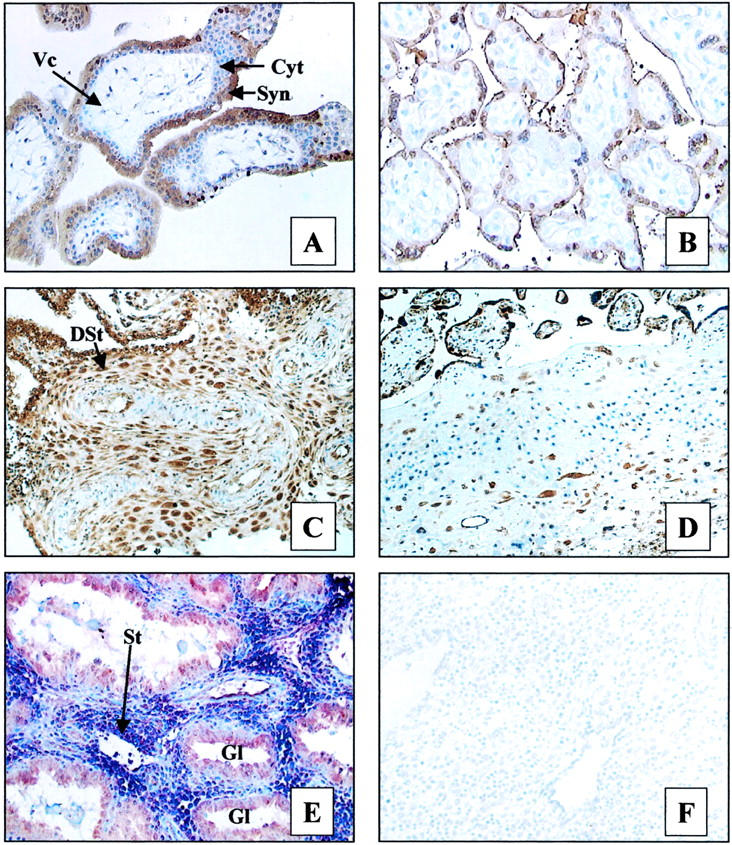
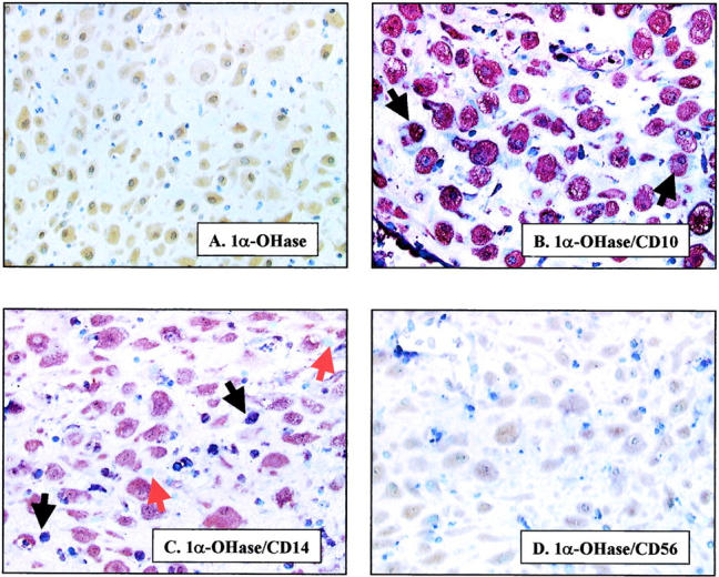
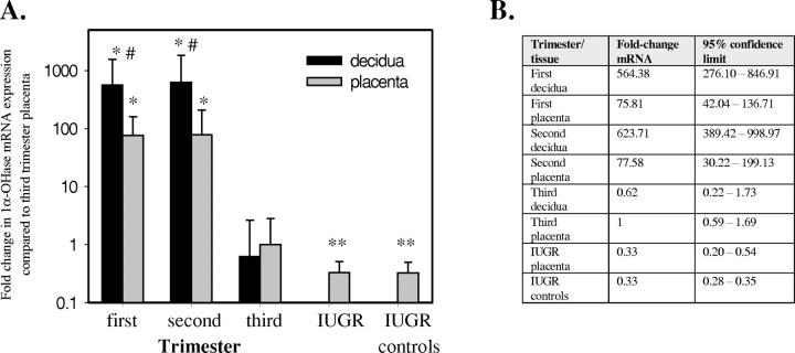
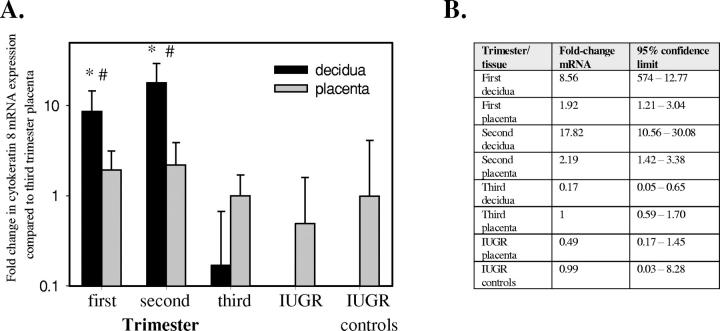
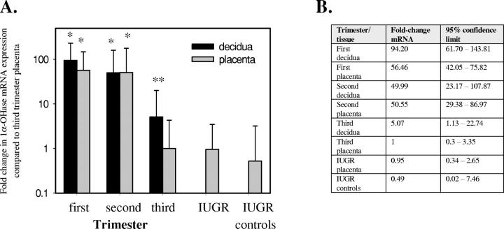
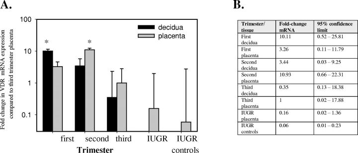
In addition to its classical calciotropic effects, the active form of vitamin D, 1,25-dihydroxyvitamin D(3) (1,25(OH)(2)D(3)) is a potent anti-proliferative/immunomodulatory secosteroid. The enzyme that catalyzes the synthesis of 1,25(OH)(2)D(3), 1alpha-hydroxylase (1alpha-OHase), is expressed in many human tissues, highlighting its possible role as an autocrine/paracrine activator of vitamin D. Immunohistochemical and RNA analyses were used to characterize the ontogeny of 1alpha-OHase expression in human placenta and decidua. Protein for 1alpha-OHase was detectable in trophoblast and decidua; the latter being stronger in decidualized stromal cells than macrophages, with no staining of lymphocytes. Quantitative reverse transcriptase-polymerase chain reaction was used to assess changes in mRNA expression for 1alpha-OHase at different gestations: first (mean, 9.1 +/- 1.5 weeks); second (mean, 14 +/- 1.8 weeks), and third trimester (mean, 39.3 +/- 2.5 weeks). 1alpha-OHase expression in decidua was approximately 1000-fold higher in first (95% confidence limits, 611 to 1376) and second (95% confidence limits, 633 to 1623) trimester biopsies when compared with the third trimester (95% confidence limits, 0.36 to 2.81) (both P < 0.001). In placenta, 1alpha-OHase expression was 80-fold higher in the first (range, 42 to 137) and second (range, 30 to 199) trimester when compared with third trimester biopsies (0.6 to 1.6) (both P < 0.001). Similar results were obtained by semiquantitative IHC. Parallel analysis of the receptor for 1,25(OH)(2)D(3) (vitamin D receptor) indicated that, as with 1alpha-OHase, highest levels of expression occurred in first trimester decidua. However, changes in vitamin D receptor mRNA expression across gestation were less pronounced than 1alpha-OHase. These spatiotemporal data emphasize the potential importance of 1alpha-OHase during early fetoplacental life and, in particular, suggest an autocrine/paracrine immunomodulatory function for the enzyme.


Figures

Figure 1.
Figure 1.
Immunohistochemical analysis of 1α-OHase expression in human placenta and decidua (brown staining). A: First trimester placenta. B: Third trimester placenta. C: First trimester decidua. D: Third trimester decidua. E: Double labeling of decidua with 1α-OHase antiserum (brown staining) and antibody to the stromal cell marker CD10 (blue staining). F: Negative control for specificity of 1α-OHase expression (preabsorption of antiserum with 200-fold excess of immunizing peptide). Tissue features: Syn, syncytiotrophoblast; Cyt, cytotrophoblast; Vc, villous core; St, stromal cells; DSt, decidual stromal cells; GL, gland. Original magnifications: ×200 (A, B, E, F); ×150 (C, D).
Figure 2.
Figure 2.
Co-localization of 1α-OHase with markers of stromal cell subtypes in first trimester decidua. A: Staining for 1α-OHase protein only (brown staining). B–D: Double labeling with 1α-OHase antiserum (brown staining) and antibodies to: stromal cell marker CD10 (blue staining) (B); monocyte marker CD14 (blue staining) (C); and lymphocyte marker CD56 (blue staining) (D). Blue staining only is shown by red arrows. Co-localization (purple staining) is shown by black arrows. Original magnifications, ×250.
Figure 3.
Figure 3.
Expression of 1α-OHase mRNA in human placenta and decidua. A: Placental and decidual 1α-OHase mRNA expression normalized for 18S rRNA levels in first, second, and third trimester samples, and placentae from patients with IUGR. All data are reported relative to a third trimester placenta value of 1. *, Significantly different from third trimester placenta, P < 0.001; **, significantly different from third trimester placenta, P < 0.05; #, significantly different from equivalent placenta, P < 0.01. B: Data expressed with 95% confidence limits. IUGR controls, gestationally matched placentae.
Figure 4.
Figure 4.
Expression of mRNA for CK-8 in human placenta and decidua. A: Placental and decidual CK-8 mRNA expression normalized for 18S rRNA levels in first, second, and third trimester samples and placentae from patients with IUGR. All data are reported relative to a third trimester placenta value of 1. *, Significantly different from third trimester placenta, P < 0.001; #, significantly different from equivalent placenta, P < 0.001. B: Data expressed with 95% confidence limits. IUGR controls, gestationally matched placentae.
Figure 5.
Figure 5.
Changes in the expression of 1α-OHase in human placenta and decidua with gestation are independent of variations in trophoblast/endometrial epithelial cell status. A: Expression of 1α-OHase mRNA in human placenta and decidua corrected for 18S rRNA and CK-8 mRNA expression. All data are reported relative to a third trimester placenta value of 1. *, Significantly different from third trimester placenta, P < 0.001; **, significantly different from third trimester placenta, P < 0.05. B: Data expressed with 95% confidence limits. IUGR controls, gestationally matched placentae.
Figure 6.
Figure 6.
Changes in the expression of VDR mRNA in human placenta and decidua with gestation. A: Expression of VDR mRNA in human placenta and decidua corrected for 18S rRNA and CK-8 mRNA expression. All data are reported relative to a third trimester placenta value of 1. *, Significantly different from third trimester placenta, P < 0.05. B: Data expressed with 95% confidence limits. IUGR controls, gestationally matched placentae.

References

    1. Feldman D: Vitamin D, parathyroid hormone, and calcium: a complex regulatory network. Am J Med 1999, 107:637-639 - PubMed
    1. Hewison M, Gacad MA, Lemire J, Adams JS: Vitamin D as a cytokine and hematopoietic factor. Rev Endocrinol Metab Disorders 2001, 2:217-227 - PubMed
    1. Jones G, Strugnell SA, DeLuca HF: Current understanding of the molecular actions of vitamin D. Physiol Rev 1998, 78:1193-1231 - PubMed
    1. Bouillon R, Garmyn M, Verstuyft A, Segaert S, Casteels K, Mathieu C: Paracrine role for calcitriol in the immune system and skin creates new therapeutic possibilities for vitamin D analogs. Eur J Endo 1995, 133:7-16 - PubMed
    1. Zehnder D, Bland R, Walker EA, Bradwell AR, Howie AJ, Hewison M, Stewart PM: Expression of 25-hydroxyvitamin D3-1α-hydroxylase in the human kidney. J Am Soc Nephrol 1999, 10:2465-2473 - PubMed

Publication types

Substances

LinkOut - more resources