. 2002 Jul;161(1):155-61.
doi: 10.1016/S0002-9440(10)64167-3.

STAT-3 overexpression and p21 up-regulation accompany impaired regeneration of fatty livers

Affiliations

STAT-3 overexpression and p21 up-regulation accompany impaired regeneration of fatty livers

Michael Torbenson et al. Am J Pathol. 2002 Jul.

Abstract

Fatty liver is an important cause of morbidity in humans and is linked to impaired liver regeneration after liver injury, but the mechanisms for impaired liver regeneration remain unknown. In the normal liver, the interleukin (IL)-6/STAT-3 pathway is thought to play a central role in regeneration because this pathway is disrupted in IL-6-deficient mice that exhibit impaired liver regeneration after 70% partial hepatectomy (PH). To determine whether inhibition of STAT-3 is involved in fatty liver-related mitoinhibition, regenerative induction of STAT-3 was compared in normal mice and leptin-deficient ob/ob mice that have fatty livers and markedly impaired liver regeneration after PH. In both groups, two waves of STAT-3 activation were observed, the first in endothelia and the second in hepatocytes. Before PH, a significantly higher percentage of ob/ob endothelial and hepatocyte nuclei expressed phosphorylated (activated) STAT-3. After PH, phospho-STAT-3 accumulated in liver nuclei of lean mice and this response was markedly exaggerated in ob/ob mice. Moreover, a striking inverse correlation was noted between hepatocyte nuclear accumulation of phospho-STAT-3 and DNA synthesis (as assessed by bromodeoxyuridine labeling), as well as cyclin D1 mRNA induction and protein expression. In contrast, STAT-3 activation was positively correlated with p21 protein expression in both groups of mice. Because these results link exaggerated STAT-3 activation with impaired hepatocyte proliferation, STAT-3 inhibition cannot be a growth-arrest mechanism in ob/ob fatty livers. Rather, hyperinduction of this factor may promote mitoinhibition by up-regulating mechanisms that impede cell cycle progression.


Figures

Figure 1.
Figure 1.
STAT-3 activation after PH. Nuclear extracts from four lean and four ob/ob mice were examined for total and phosphorylated STAT-3 with an immunoblot assay using 40 μg of nuclear proteins. A representative immunoblot is shown.
Figure 2.
Figure 2.
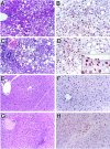
ob/ob mice showed significant steatosis at time 0 hours (A). Nuclear staining for pSTAT-3 is detected at time 0 hours in an ob/ob mouse liver (B). Twenty-four hours after PH, the ob/ob mice showed a similar degree of steatosis (C) and significantly more hepatocyte nuclear labeling for pSTAT-3 (D). Endothelial nuclear labeling can also be seen in a central vein (D). In contrast, livers from lean littermates showed no steatosis at time 0 hours (E) and were generally negative for nuclear pSTAT-3 staining (F). After PH, there were no morphological changes (G) and only occasional scattered hepatocyte nuclei were positive for pSTAT-3 (H).
Figure 3.
Figure 3.
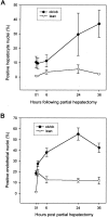
Both hepatocytes (A) and endothelial cells (B) in ob/ob mice showed increased pSTAT-3 nuclear labeling at baseline. In response to PH, the lean mice showed a modest increase in pSTAT-3 labeling that peaked at 24 hours, whereas the ob/ob mice showed a delayed, but much greater increase in nuclear staining that had not peaked by 36 hours (A). Similarly, the endothelial cells in the ob/ob mice showed increased pSTAT-3 labeling and had a delayed peak compared to the lean mice. Five hundred hepatocytes and 200 endothelial cells (100 portal vein, 100 central vein) were scored in each liver at every time point. Each time point represents two to three mice. Results are shown as mean ± SEM.
Figure 4.
Figure 4.
Hepatocyte nuclear incorporation of BrdU was impaired in ob/ob mice after PH. Mice had BrdU injected into the peritoneal cavity 2 hours before sacrifice. Results are for two to three mice per group (mean ± SEM).
Figure 5.
Figure 5.
Cyclin D1 mRNA (A) and protein levels (B) were lower in ob/ob mice than lean mice after PH. A: Changes in the mRNA levels were measured using an RNase protection assay. 18S RNA was concurrently evaluated to ensure lane-to-lane equivalency of RNA loading and transfer and a representative autoradiograph is shown. The graph summarizes the data with two to three mice per time point. B: Cyclin D1 protein levels were evaluated with an immunoblot assay using 40 μg per lane. A representative blot is shown. Results are for two to three mice per group (mean ± SEM), except for ob/ob at 36 hours, which is a single mouse.
Figure 6.
Figure 6.
CKIs p21 and p27 levels were higher at baseline and all subsequent time points in ob/ob mice compared to lean littermates. p21 levels increased in both ob/ob and lean mice, whereas p27 levels showed an initial decrease after PH followed by a modest increase. p21 and p27 protein levels were evaluated with an immunoblot assay using 40 μg per lane. A representative blot is shown. The graph summarizes the data with two to three mice per time point. Results are for two to three mice per group (mean ± SEM).

References

    1. Ludwig J, Viggiano TR, McGill DB, Oh BJ: Nonalcoholic steatohepatitis: Mayo Clinic experiences with a hitherto unnamed disease. Mayo Clin Proc 1980, 55:434-438 - PubMed
    1. Wanless IR, Lentz JS: Fatty liver hepatitis (steatohepatitis) and obesity: an autopsy study with analysis of risk factors. Hepatology 1990, 12:1106-1110 - PubMed
    1. Lee RG: Nonalcoholic steatohepatitis: a study of 49 patients. Hum Pathol 1989, 20:594-598 - PubMed
    1. Lee RG: Diagnostic Liver Pathology 1994. Mosby, St. Louis
    1. Lindblad A, Glaumann H, Strandvik B: Natural history of liver disease in cystic fibrosis. Hepatology 1999, 30:1151-1158 - PubMed

Publication types

MeSH terms