. 2002 Jul;161(1):173-82.
doi: 10.1016/S0002-9440(10)64169-7.

Terminal bronchioles harbor a unique airway stem cell population that localizes to the bronchoalveolar duct junction

Affiliations

Terminal bronchioles harbor a unique airway stem cell population that localizes to the bronchoalveolar duct junction

Adam Giangreco et al. Am J Pathol. 2002 Jul.

Abstract

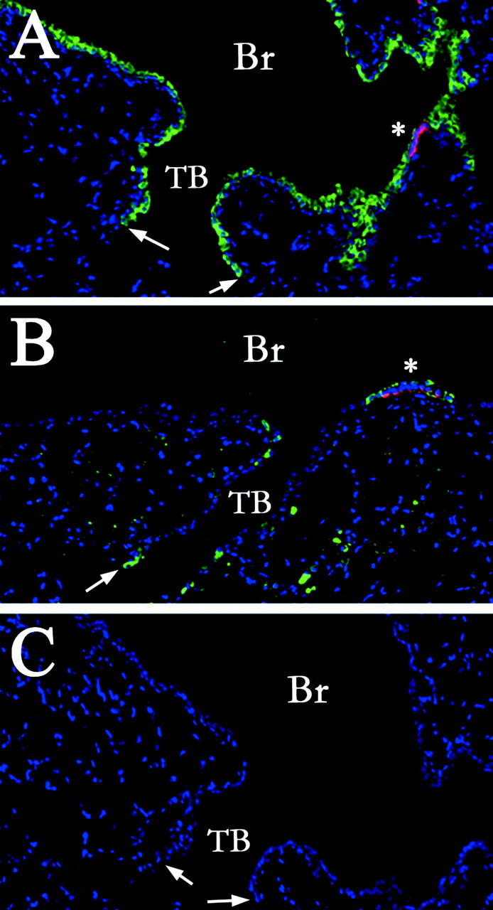
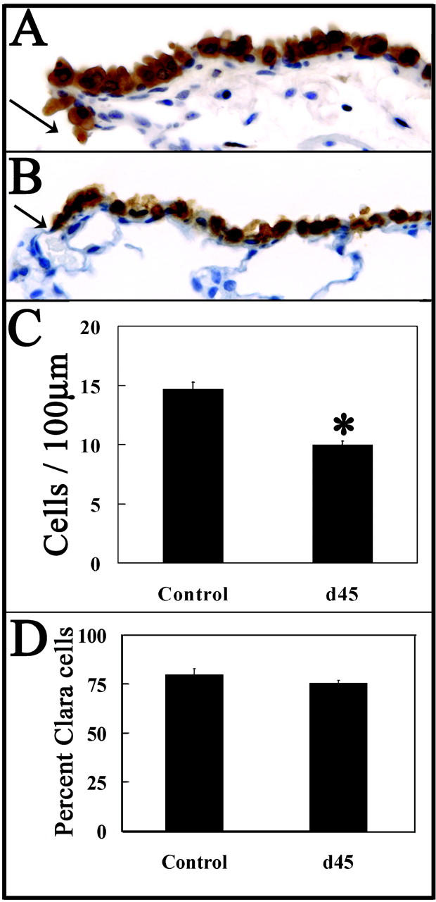
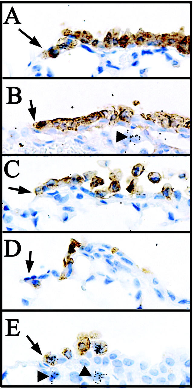
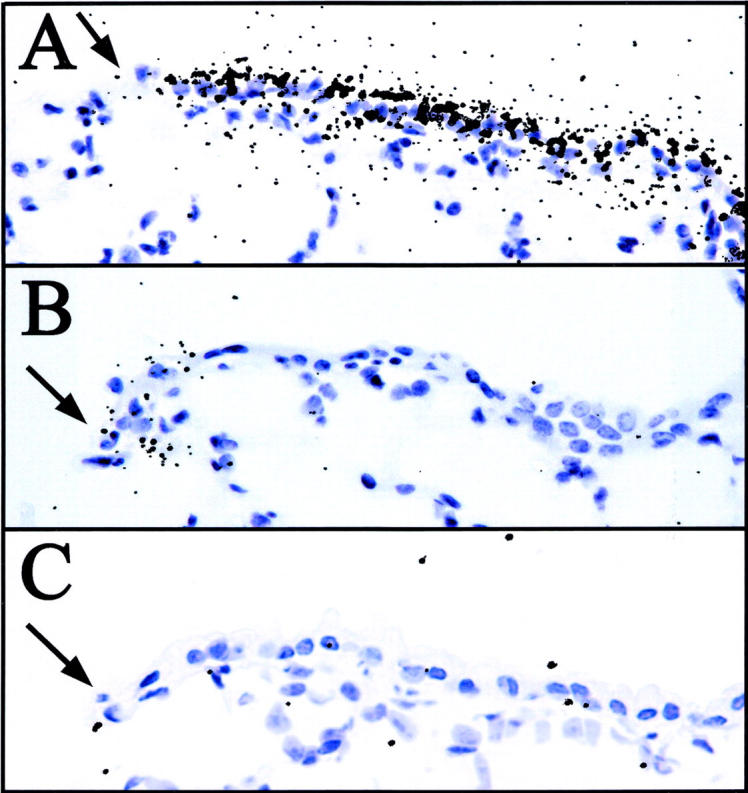
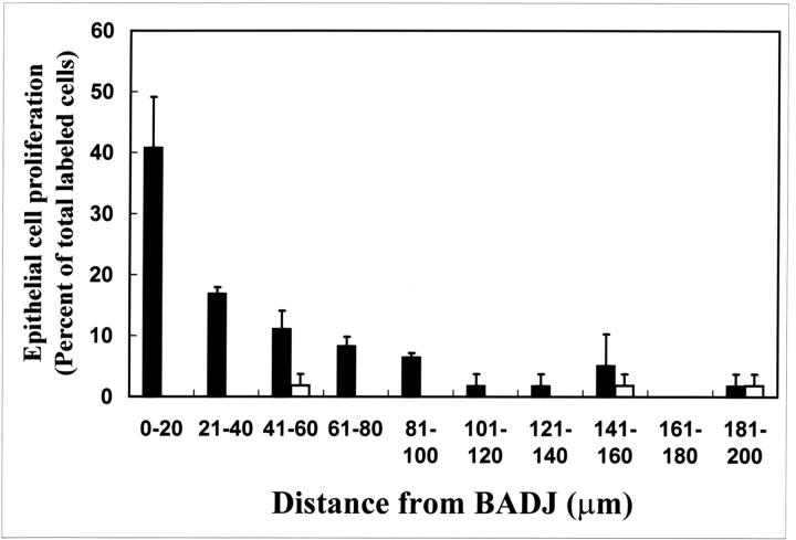
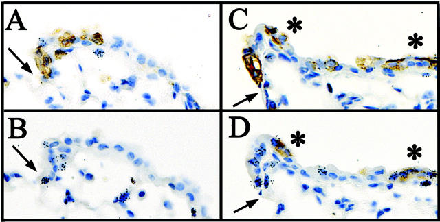
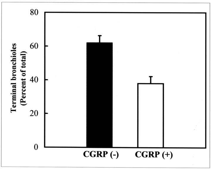
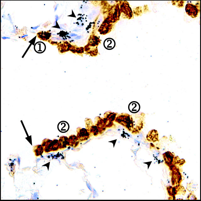
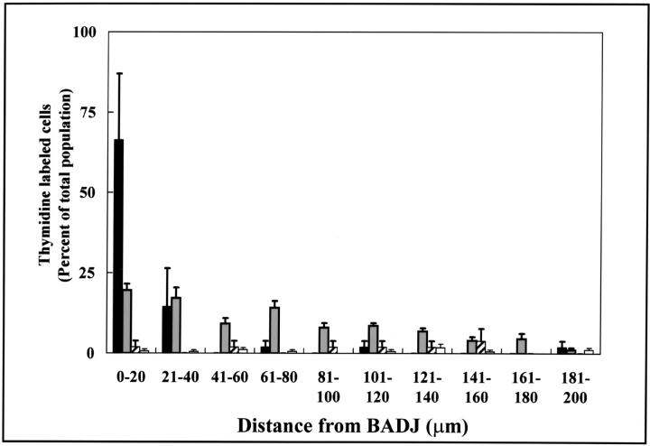
Cellular mechanisms contributing to renewal of terminal bronchioles remain poorly defined. Our previous studies identified pollutant-resistant Clara cell secretory protein (CCSP)-expressing stem cells that localize to the neuroepithelial body (NEB) and contribute to renewal of the proximal bronchiolar epithelium. However, activation of NEB-associated stem cells is unlikely to contribute to renewal of terminal bronchiolar epithelium because of the paucity of NEBs at this location. Goals of this study were to determine the location and properties of cells contributing to renewal of terminal bronchioles after Clara cell depletion. Pollutant-resistant CCSP-expressing cells were identified that localized to the bronchoalveolar duct junction (BADJ) and contribute to restoration of a phenotypically diverse epithelium. CCSP-expressing cells comprise the predominant proliferative population in initial terminal bronchiolar repair and include a population of label-retaining cells suggesting that they maintain characteristics of a stem cell population. Furthermore, immunohistochemical co-localization studies involving CCSP and the NEB-specific marker calcitonin gene-related peptide indicate that BADJ-associated CCSP-expressing stem cells function independently of NEB microenvironments. These studies identify a BADJ-associated, NEB-independent, CCSP-expressing stem cell population in terminal bronchioles and support the notion that regiospecific stem cell niches function to maintain epithelial diversity after injury.


Figures

Figure 1.
Figure 1.
Pollutant-resistant CE cells occupy multiple distinct microenvironments within the conducting airway epithelium. CCSP (green)- and CGRP (red)-immunopositive cells were detected by dual-color immunofluorescent staining of lung tissue sections from either untreated control mice (A) or mice treated with 275 mg/kg of naphthalene and recovered for 24 hours (B). Primary antibodies against CCSP and CGRP were omitted from untreated lung samples as a control for antibody specificity (C). Nuclei were counterstained with 4,6-diamidino-2-phenylindole (blue fluorescence, A–C). Immunofluorescent cellular debris (lacking nuclei) in the alveolar space (B) is an artifact of inflation fixation. Analysis revealed a significant decrease in the number of CCSP-positive cells in both the bronchiole (Br) and terminal bronchiolar (TB) airways of naphthalene-treated mice (B). Rare populations of naphthalene-resistant CCSP-immunoreactive cells were located directly adjacent to CGRP-positive NEBs (A and B, asterisks), or at the BADJ (A and B, arrows). Original magnifications, ×100.
Figure 2.
Figure 2.
Cells within terminal bronchioles support regeneration of airways of appropriate cellular diversity. CCSP-immunopositive cells in terminal bronchioles were detected by immunohistochemistry 45 days after intraperitoneal injection of vehicle (A) or 275 mg/kg of naphthalene (B). Nuclei were counterstained with hematoxylin; arrows indicate the BADJ (A and B). Morphometric analyses of cell density (total cells/100 μm basement membrane, C), and representation of Clara cells (percent Clara cells/total epithelial cells, D) were performed as described in Materials and Methods. Eight to 16 terminal bronchioles were analyzed per lung (n = lungs from three mice). Significant changes in overall cell density (C, ✻, P < 0.005) but not percentage of Clara cells (D) indicates an appropriate differentiation potential of epithelial cells within terminal bronchioles after naphthalene-induced injury. Original magnifications, ×400.
Figure 3.
Figure 3.
CE cells adjacent to the BADJ are pollutant-resistant and represent the initial proliferative epithelial population. Mice were exposed to 275 mg/kg of naphthalene and recovered for 0, 6, 12, 24, and 36 hours (A to E, respectively). Proliferating mitotic cells were labeled with 2.5 mCi of [3H]-TdR/kg body weight 1 hour before sacrifice. CE cells were detected by immunohistochemistry using diaminobenzidine detection (brown) and sites of [3H]-TdR incorporation identified by autoradiography (black grains). Nuclei were counterstained with hematoxylin. Naphthalene-resistant CE cells are detectable adjacent to the BADJ at all time points (A–E, arrows), and are the initial proliferative epithelial cell type 36 hours after naphthalene administration (E, black grains). Arrowheads in B and E indicate underlying proliferative fibroblasts. Original magnifications, ×400.
Figure 4.
Figure 4.
BADJ-associated pollutant-resistant cells maintain CCSP gene expression. CCSP gene expression in untreated control (A) and during the period of maximum injury 24 hours after 275 mg/kg of naphthalene treatment (B) was determined by in situ hybridization using an anti-sense [35S]-labeled CCSP riboprobe. Untreated, CCSP−/− tissue was used as a negative control (C). Hybridization was detected by autoradiography (black grains), and indicates a population of pollutant-resistant CCSP mRNA-positive cells immediately adjacent to the BADJ (A–C, arrows). Panels are representative of terminal bronchioles of three animals per exposure group. Original magnifications, ×400.
Figure 5.
Figure 5.
Proliferative CE cells are preferentially associated with the BADJ. Distribution of [3H]-TdR-labeled cells was determined within the terminal bronchiole 36 hours after 275 mg/kg of naphthalene exposure. Terminal bronchioles were divided into 20-μm segments originating from the BADJ and the number of labeled CE) (black bars) or CCSP-negative (white bars) cells in each segment enumerated. Fifty-seven percent of labeled CE cells were found within 40 μm (6 cell diameters) of the BADJ. In contrast, CCSP-negative cells contribute only slightly to this initial proliferative population and were not spatially restricted within the terminal bronchiole. Data represent the summation of 8 to 10 terminal bronchioles/section (n = 3 animals) ± SEM.
Figure 6.
Figure 6.
Proliferative CE cell populations function independently of PNECs/NEBs in the terminal bronchiole. Adjacent serial section immunostaining for CCSP (A and C) and CGRP (B and D) was coupled with autoradiography (black grains) to determine whether terminal bronchiolar proliferation was preferentially associated with CGRP-immunopositive PNECs/NEBs. The majority (75%) of terminal bronchioles contain numerous mitotic CE cells adjacent to the BADJ (A) yet lack any CGRP-positive PNECs/NEBs in the adjacent serial section (B) 36 hours after 275 mg/kg of naphthalene. A minority of terminal bronchioles (25%) appear to contain both CCSP (C)- and CGRP (D)-positive mitotic cells by adjacent serial section analysis. Interestingly, the occurrence of NEBs (C and D, asterisks) appeared to relax the requirement for BADJ association among proliferating CE cells of the terminal bronchiole (black grains; A and B versus C and D).
Figure 7.
Figure 7.
The majority of terminal bronchioles lack PNECs/NEBs after naphthalene injury. Dual-color immunofluorescent staining for CCSP and CGRP proteins was used to co-localize CE and PNEC populations within terminal bronchioles 24 hours after exposure to 275 mg/kg of naphthalene. Twenty consecutive 5-μm-adjacent serial lung sections were stained for the presence of CCSP and CGRP and all terminal bronchioles containing a visible BADJ were analyzed (minimum of five terminal bronchioles/animal; n = 5 animals). Although all terminal bronchioles analyzed contained detectable CCSP-immunoreactive cells at or near the BADJ, 62 ± 5% lacked any CGRP-positive PNECs/NEBs at any location within the terminal bronchiole. Error bars represent SEM.
Figure 8.
Figure 8.
BADJ-associated CE cells represent an infrequently cycling (label-retaining) cell population. Mice were exposed to 275 mg/kg of naphthalene and [3H]-TdR was administered continuously for the subsequent 7 day period via a miniosmotic pump. Recovering mice were sacrificed 45 days after naphthalene exposure, at which time CCSP protein expression and label retention was visualized by immunohistochemistry and autoradiography of lung tissue sections. The majority of labeled cells contained an average of 5 to 10 grains/nuclei, and were composed of both CCSP-immunopositive (circled 2) and -immunonegative cells (not evident in field). A rare population of CE cells located near the BADJ (arrow) were more heavily labeled with an average of >15 grains/nuclei (circled 1). Fibroblasts, identified by location and nuclear morphology, also contained large numbers of grains/nuclei (arrowheads). Original magnification, ×1000.
Figure 9.
Figure 9.
CE label-retaining cells are preferentially located at the BADJ. The distribution of [3H]-TdR-labeled (5 to 10 grains/nuclei) and label-retaining (>15 grains/nuclei) cells in terminal bronchioles of animals exposed to 275 mg/kg of naphthalene and recovered for 45 days were assessed by morphometric analysis. The percentage of each thymidine-labeled cell population was plotted independently as a function of distance in 20-μm increments originating from the BADJ. A correlation was observed between proximity to the BADJ and the abundance of CE, label-retaining cell populations (black bars). Very few CCSP-negative cells label-retaining cells (hatched white bars) were identified within terminal bronchioles, and these cells were not associated with the BADJ. Both CCSP-immunopositive (gray bars) and -negative (white bars) labeled (5 to 10 grains/nuclei) cell populations were distributed randomly throughout the terminal bronchiole. Data are the summation of 23 terminal bronchioles (n = 4 mice).

Similar articles

Cited by

References

    1. Barth PJ, Wolf M, Ramaswamy A: Distribution and number of Clara cells in the normal and disturbed development of the human fetal lung. Pediatr Pathol 1994, 4:637-651 - PubMed
    1. Plopper CG, Mariassy AT, Hill LH: Ultrastructure of the nonciliated bronchiolar epithelial (Clara) cell of mammalian lung: I. A comparison of rabbit, guinea pig, rat, hamster, and mouse. Exp Lung Res 1980, 1:139-154 - PubMed
    1. Plopper CG, Mariassy AT, Hill LH: Ultrastructure of the nonciliated bronchiolar epithelial (Clara) cell of mammalian lung: II. A comparison of horse, steer, sheep, dog, and cat. Exp Lung Res 1980, 1:155-169 - PubMed
    1. Plopper CG, Hill LH, Mariassy AT: Ultrastructure of the nonciliated bronchiolar epithelial (Clara) cell of mammalian lung. III. A study of man with comparison of 15 mammalian species. Exp Lung Res 1980, 1:171-180 - PubMed
    1. Plopper CG, Mariassy AT, Wilson DW, Alley JL, Nishio SJ, Nettesheim P: Comparison of nonciliated tracheal epithelial cells in six mammalian species: ultrastructure and population densities. Exp Lung Res 1983, 5:281-294 - PubMed

Publication types

MeSH terms