

Eosinophil recruitment in type-2 hypersensitivity pulmonary granulomas: source and contribution of monocyte chemotactic protein-3 (CCL7)
- PMID: 12107110
- PMCID: PMC1850678
- DOI: 10.1016/S0002-9440(10)64177-6
Eosinophil recruitment in type-2 hypersensitivity pulmonary granulomas: source and contribution of monocyte chemotactic protein-3 (CCL7)
Abstract
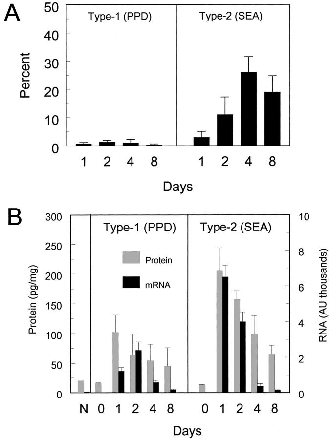
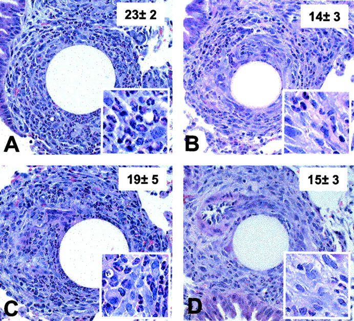
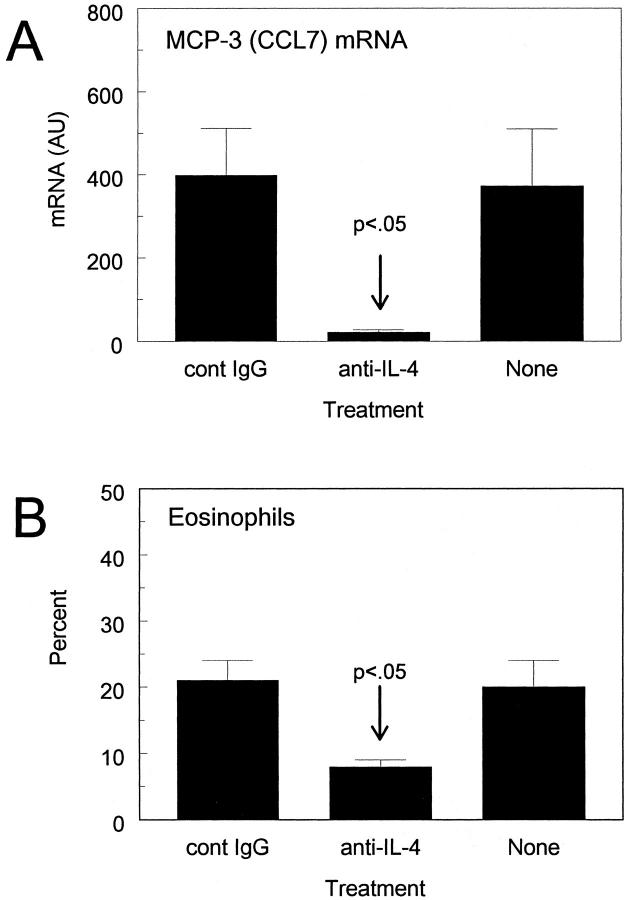
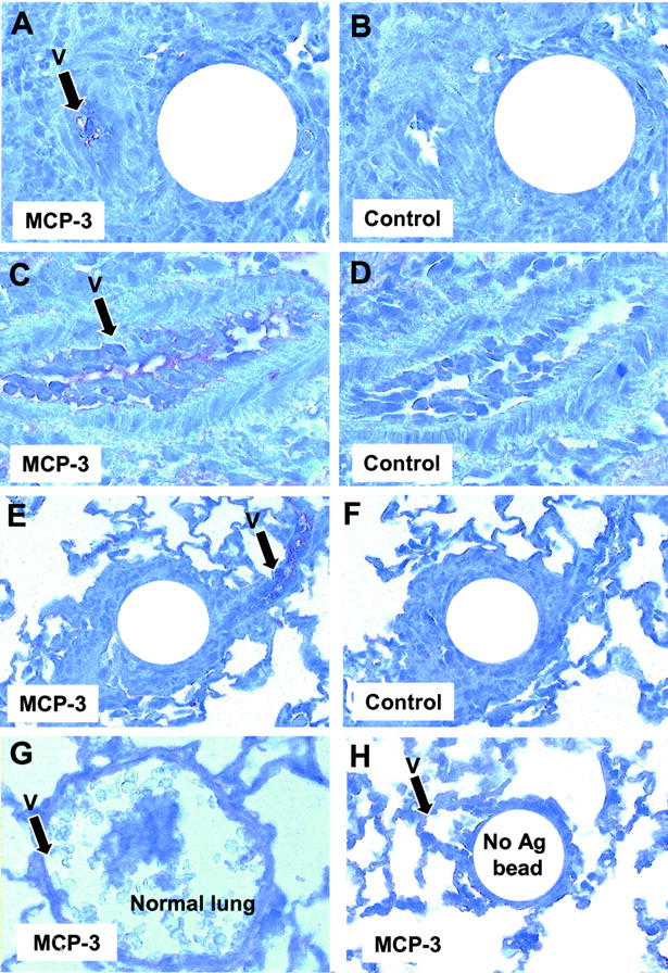
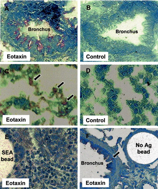
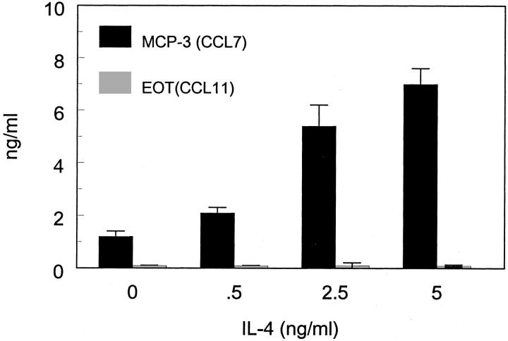
Monocyte chemotactic protein-3 (MCP-3/CCL7) has potent eosinophil chemoattractant properties. The present study determined its relative contribution to the formation of Th2 cytokine-mediated (type-2) eosinophil-rich interstitial lung granulomas induced by antigens of Schistosoma mansoni eggs. Both MCP-3 transcripts and protein levels were more strongly expressed in lungs with type-2 than with type-1 (mycobacterial antigen-elicited Th1-mediated) granulomas. In vivo treatment with neutralizing antibodies demonstrated that MCP-3 abrogated eosinophil accumulation in type-2 lesions by 40 to 50%. Immunohistochemical staining revealed that MCP-3 localized to vessels in or near granulomas suggesting that endothelial cells were an important in situ source of MCP-3. Maximal MCP-3 transcript expression was abrogated by anti-interleukin-4 treatment. Furthermore, cultured mouse lung endothelial cells displayed augmented MCP-3 production in response to interleukin-4. Together, these results suggest that MCP-3 contributes to a significant component of eosinophil recruitment in the type-2 interstitial granuloma formation and Th2 cytokines promote its production.
Figures
Similar articles
-
Interleukin 4 and 13 participation in mycobacterial (type-1) and schistosomal (type-2) antigen-elicited pulmonary granuloma formation: multiparameter analysis of cellular recruitment, chemokine expression and cytokine networks.Cytokine. 2000 May;12(5):432-44. doi: 10.1006/cyto.1999.0595. Cytokine. 2000. PMID: 10857756
-
Chemokine responses in schistosomal antigen-elicited granuloma formation.Parasite Immunol. 2002 Jun;24(6):285-94. doi: 10.1046/j.1365-3024.2002.00466.x. Parasite Immunol. 2002. PMID: 12102713
-
Role of monocyte chemoattractant protein-1 (MCP-1) in Th1 (mycobacterial) and Th2 (schistosomal) antigen-induced granuloma formation: relationship to local inflammation, Th cell expression, and IL-12 production.J Immunol. 1996 Nov 15;157(10):4602-8. J Immunol. 1996. PMID: 8906839
-
AMD3465, a novel CXCR4 receptor antagonist, abrogates schistosomal antigen-elicited (type-2) pulmonary granuloma formation.Am J Pathol. 2006 Aug;169(2):424-32. doi: 10.2353/ajpath.2006.051234. Am J Pathol. 2006. PMID: 16877345 Free PMC article.
-
Mechanisms regulating eosinophil extravasation in asthma.Eur Respir J Suppl. 1996 Aug;22:136s-140s. Eur Respir J Suppl. 1996. PMID: 8871059 Review.
Cited by
-
Establishment of experimental eosinophilic vasculitis by IgE-mediated cutaneous reverse passive arthus reaction.Am J Pathol. 2009 Jun;174(6):2225-33. doi: 10.2353/ajpath.2009.080223. Epub 2009 Apr 23. Am J Pathol. 2009. PMID: 19389931 Free PMC article.
-
Fibrogenic and redox-related but not proinflammatory genes are upregulated in Lewis rat model of chronic silicosis.J Toxicol Environ Health A. 2011;74(19):1261-79. doi: 10.1080/15287394.2011.595669. J Toxicol Environ Health A. 2011. PMID: 21830856 Free PMC article.
-
Urokinase-deficient mice fail to generate a type 2 immune response following schistosomal antigen challenge.Infect Immun. 2004 Jan;72(1):461-7. doi: 10.1128/IAI.72.1.461-467.2004. Infect Immun. 2004. PMID: 14688127 Free PMC article.
-
Protection Elicited by Attenuated Live Yersinia pestis Vaccine Strains against Lethal Infection with Virulent Y. pestis.Vaccines (Basel). 2021 Feb 16;9(2):161. doi: 10.3390/vaccines9020161. Vaccines (Basel). 2021. PMID: 33669472 Free PMC article.
-
Therapeutic Targets in Allergic Conjunctivitis.Pharmaceuticals (Basel). 2022 Apr 28;15(5):547. doi: 10.3390/ph15050547. Pharmaceuticals (Basel). 2022. PMID: 35631374 Free PMC article. Review.
References
-
- Wardlaw AJ, Brightling C, Green R, Woltmann G, Pavord I: Eosinophils in asthma and other allergic diseases. Br Med Bull 2000, 56:985-1003 - PubMed
-
- Gao JL, Sen AI, Kitaura M, Yoshie O, Rothenberg ME, Murphy PM, Luster AD: Identification of a mouse eosinophil receptor for the CC chemokine eotaxin. Biochem Biophys Res Commun 1996, 223:679-684 - PubMed
-
- White JR, Imburgia C, Dul E, Appelbaum E, O’Donnell K, O’Shannessy DJ, Brawner M, Fornwald J, Adamou J, Elshourbagy NA, Kaiser K, Foley JJ, Schmidt DB, Johanson K, Macphee C, Moores K, McNulty D, Scott GF, Schleimer RP, Sarau HM: Cloning and functional characterization of a novel human CC chemokine that binds to the CCR3 receptor and activates human eosinophils. J Leukoc Biol 1997, 62:667-675 - PubMed
-
- Grimaldi JC, Yu NX, Grunig G, Seymour BWP, Cottrez F, Robinson DS, Hosken N, Ferlin WG, Wu XY, Soto H, O’Garra A, Howard MC, Coffman RL: Depletion of eosinophils in mice through the use of antibodies specific for C-C chemokine receptor 3 (CCR3). J Leukoc Biol 1999, 65:846-853 - PubMed
Publication types
MeSH terms
Substances
Grants and funding
LinkOut - more resources
Full Text Sources
Medical
Miscellaneous
