. 2002 Jul;161(1):291-300.
doi: 10.1016/S0002-9440(10)64181-8.

Complicated mechanisms of class II transactivator transcription deficiency in small cell lung cancer and neuroblastoma

Affiliations

Complicated mechanisms of class II transactivator transcription deficiency in small cell lung cancer and neuroblastoma

Takuya Yazawa et al. Am J Pathol. 2002 Jul.

Abstract

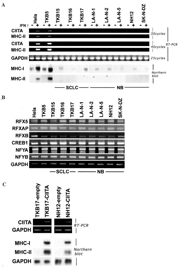
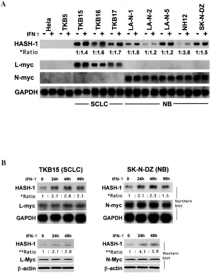
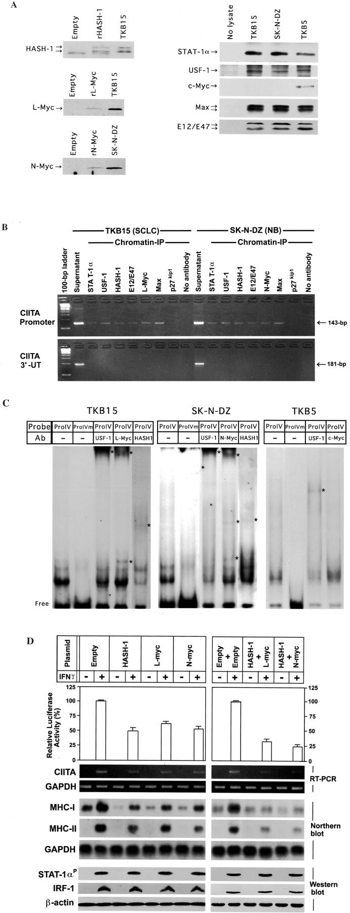
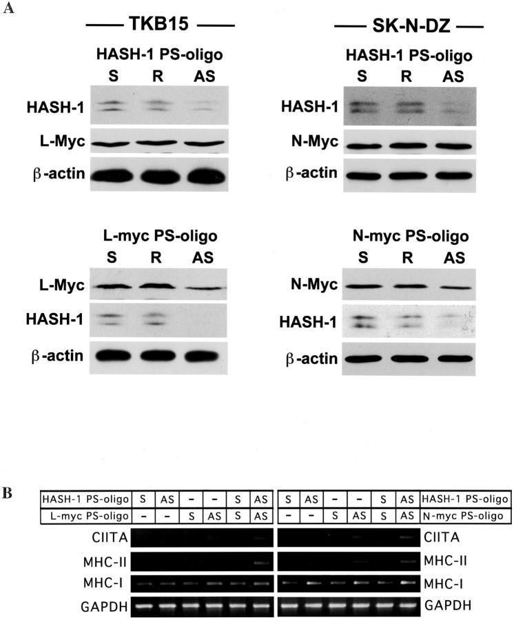
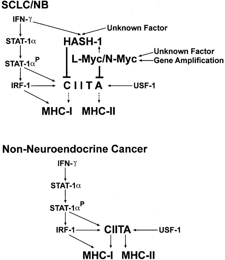
Small cell lung cancer (SCLC) and neuroblastoma (NB), the most aggressive adult and infant neuroendocrine cancers, respectively, are immunologically characterized by a severe reduction in major histocompatibility complex (MHC) that is indispensable for anti-tumor immunity. We had reported that the severe reduction of MHC in SCLC was caused by a deficient interferon (IFN)-gamma-inducible expression of class II transactivator (CIITA) that is known as a very important transcription factor for IFN-gamma-inducible class II and class I MHC expression (Yazawa T, Kamma H, Fujiwara M, Matsui M, Horiguchi H, Satoh H, Fujimoto M, Yokohama K, Ogata T: Lack of class II transactivator causes severe deficiency of HLA-DR expression in small cell lung cancer. J Pathol 1999, 187:191-199). Here, we demonstrate that the reduction of MHC in NB was also caused by a deficient IFN-gamma-inducible expression of CIITA and that the deficiency in SCLC and NB was caused by similar mechanisms. Human achaete-scute complex homologue (HASH)-1, L-myc, and N-myc, which are specifically overexpressed in SCLC and NB, bound to the E-box in CIITA promoter IV and reduced the transcriptional activity. Anti-sense oligonucleotide experiments revealed that overexpressed L-myc and N-myc lie upstream in the regulatory pathway of HASH-1 expression. The expression of HASH-1 was also up-regulated by IFN-gamma. Our results suggest that SCLC and NB have complicated mechanisms of IFN-gamma-inducible CIITA transcription deficiency through the overexpressed HASH-1, L-myc, and N-myc. These complicated mechanisms may play an important role in the escape from anti-tumor immunity.


Figures

Figure 1.
Figure 1.
Severe deficiency of constitutive and IFN-γ-inducible CIITA/MHC expression in SCLC and NB. A: RT-PCR and Northern blot analyses on constitutive and IFN-γ-inducible expression of CIITA, MHC-I, and MHC-II mRNA. The IFN-γ induction of CIITA and MHC-II is extremely deficient in SCLC and NB cell lines. Both constitutive and IFN-γ-inducible MHC-I expression is also significantly lower in SCLC and NB cell lines than in nonneuroendocrine cancers. B: RT-PCR analyses on MHC-II-specific transcription factors. The other transcription factors crucial for MHC-II, RFX5, RFXAP, RFXB, CREB1, and NFY are constitutively expressed well in SCLC, NB, and nonneuroendocrine cancer cell lines. C: RT-PCR and Northern blot analyses on CIITA and MHC expression in empty vector-transfected (TKB17-empty, NH12-empty) or CIITA-transfected (TKB17-CIITA, NH12-CIITA) cells. MHC-I and MHC-II expression are improved through the CIITA transfection both in TKB17 (SCLC) and NH12 (NB).
Figure 2.
Figure 2.
A: Northern blot analysis on the expression and alteration by IFN-γ treatment of HASH-1, L-myc, and N-myc. All SCLC and NB cell lines express HASH-1, and IFN-γ treatment up-regulates the HASH-1 expression. The ratio of HASH-1 mRNA expression (asterisk) was calculated as follows: [density of HASH-1 mRNA (IFN-γ(+))/density of GAPDH mRNA (IFN-γ(+))]/[density of HASH-1 mRNA (IFN-γ(−))/density of GAPDH mRNA (IFN-γ(−))]. L-myc is overexpressed in SCLC cell lines and N-myc is overexpressed in NB cell lines, whereas nonneuroendocrine cancers, HeLa and TKB5, do not express HASH-1, L-myc, or N-myc. The expression levels of L-myc and N-myc do not alter with IFN-γ treatment. B: Alteration of HASH-1 mRNA and protein expression in TKB15 (SCLC) and SK-N-DZ (NB) through IFN-γ treatment. The ratio of HASH-1 mRNA level (asterisk) was calculated as follows: [density of HASH-1 mRNA (IFN-γ(+))/density of GAPDH mRNA (IFN-γ(+))]/[density of HASH-1 mRNA (IFN-γ(−))/density of GAPDH mRNA (IFN-γ(−))]. The ratio of HASH-1 protein expression (double asterisk) was calculated as follows: [density of HASH-1 protein (IFN-γ(+))/density of β-actin protein (IFN-γ(+))]/[density of HASH-1 protein (IFN-γ(−))/density of β-actin protein (IFN-γ(−))]. HASH-1 is up-regulated at both the mRNA and protein level by the IFN-γ treatment.
Figure 3.
Figure 3.
A: Expression vector quality and ability to immunoprecipitate cognate antigen that each antibody (anti-HASH-1, anti-L-Myc, anti-N-Myc, anti-STAT-1α, anti-USF-1, anti-c-Myc, Max, or anti-E12/E47 antibody) recognizes. Immunoprecipitation studies were performed using in vitro transcription and translation reactants from HASH-1-, L-Myc-, or N-Myc-encoding pZeoSV2 vector and nuclear lysate from TKB15, SK-N-DZ, or TKB5. The anti-MASH-1 antibody used in this experiment cross-reacts with recombinant HASH-1 (rHASH-1) and intrinsic HASH-1 protein. The anti-L-Myc antibody reacts with recombinant L-Myc (rL-Myc) and intrinsic L-Myc. The anti-N-Myc antibody reacts with rN-Myc as well as intrinsic N-Myc. Abundant STAT-1α, USF-1, Max, and E12/E47 proteins are immunoprecipitated from both TKB15, SK-N-DZ, and TKB5. The nuclear lysate from TKB5 contains abundant c-Myc protein. These antibodies were used in the chromatin immunoprecipitation assay. Empty: In vitro transcription and translation using a pZeoSV2-empty vector. B: Chromatin immunoprecipitation assay-based PCR using DNA fragments immunoprecipitated with the indicated antibodies. Top: PCR using CIITA promoter IV-specific primers. Bottom: PCR using CIITA 3′-untranslated region (3′-UT)-specific primers. The results show that not only essential transcription factors (STAT-1α and USF-1) but also HASH-1, L-Myc, N-Myc, and their heterodimeric partners (E12/E47 and Max) bind to the E-box in CIITA promoter IV. No primary antibody and p27kip1 antibody: negative control. Sonicated DNA: positive control. Ladder: 100-bp ladder marker. C: Electromobility shift assay using nuclear lysate from TKB15, SK-N-DZ, or TKB5. Although the signals supershifted by the supplement of anti-USF-1 antibody are found both in TKB15, SK-N-DZ, and TKB5, the patterns are different. The signals in TKB15 and SK-N-DZ are also supershifted by the supplement of anti-L-Myc, anti-N-Myc, or anti-HASH-1 antibody. A signal supershifted by the supplement of anti-c-Myc antibody is not isolated in TKB5. ProIV: CIITA promoter IV probe containing IFN-γ-activating site (GAS) and E-box. ProIVm: CIITA promoter IV probe containing GAS and mutated E-box. Ab: Antibody. Asterisk: Supershifted complexes. Free: Free probe. D: Top, Reporter gene assay using TKB5 cells into which neuroendocrine cell-specific bHLH transcription factors were stably transfected, a CIITA promoter IV-encoded luciferase reporter vector, and a control vector. TKB5 was used as a parental cell because it revealed high levels of IFN-γ-inducible CIITA and MHC expression. The reporter and control vectors were transiently co-transfected. Each transient transfectant/co-transfectant is representative of several stable clones. Values of luciferase activity are means + SD of the relative activities in three independent experiments performed in triplicate. The CIITA promoter activity of stably bHLH-transfected TKB5 cell lines is lower than that of the empty vector-transfected one. Co-transfection of HASH-1 with L-myc or N-myc further down-regulates the CIITA promoter IV activity. Bottom: RT-PCR analysis on CIITA mRNA, Northern blot analyses on MHC-I and MHC-II mRNA, and Western blot analyses on phosphorylated STAT-1α, IRF-1, and USF-1 expression in the stable transfectants/co-transfectants. CIITA, MHC-I, and MHC-II mRNA show a decrease on forced expression of HASH-1, L-myc, and N-myc, and these data support those of reporter gene assay. USF-1 is constitutively expressed and IRF-1 (IFN-γ treatment for 24 hours) and phosphorylated STAT-1α (IFN-γ treatment for 30 minutes) are induced well even in HASH-1-, L-myc-, and N-myc-transfectants, or double transfectants.
Figure 4.
Figure 4.
The overexpression of L-myc and N-myc is involved in HASH-1 expression. A: Top, Change in the expression of L-Myc and N-Myc proteins associated with forced repression of HASH-1. Bottom: Change in the expression of HASH-1 protein associated with forced repression of L-Myc or N-Myc. Although forced repression of HASH-1 does not influence the expression levels of L-Myc and N-Myc, HASH-1 is down-regulated by the forced repression of L-Myc and N-Myc. S: Sense PS-oligo treatment. R: Reverse PS-oligo treatment. AS: Anti-sense PS-oligo treatment. B: Increase of IFN-γ-inducible CIITA and MHC mRNA expression through forced repression of HASH-1, L-Myc, and N-Myc. The treatment with the anti-sense L-myc- or N-myc-PS-oligo alone as well as anti-sense HASH-1-PS-oligo alone somewhat increases the IFN-γ-inducible expression of CIITA and MHC. The IFN-γ-inducible CIITA and MHC expression are more improved through the double anti-sense PS-oligo treatments (HASH-1 AS + L-Myc AS or HASH-1 AS + N-Myc AS).
Figure 5.
Figure 5.
A scheme for the mechanisms of severe CIITA deficiency in SCLC and NB. In nonneuroendocrine cancers, the CIITA gene is transactivated through the phosphorylated STAT-1α, IRF-1, and USF-1, and this is followed by transactivation of MHC-II and CIITA-mediated MHC-I expression. In addition, the IFN-stimulated response element (ISRE)-mediated MHC-I activation is generated by IRF-1. However, in SCLC and NB, the IFN-γ-inducible CIITA expression is severely repressed by overexpressed HASH-1, L-myc, and N-myc. The IFN-γ treatment up-regulates the HASH-1 expression in SCLC and NB. The overexpressed L-myc and N-myc up-regulates the HASH-1 expression in SCLC and NB.

Comment in

Similar articles

Cited by

References

    1. Travis WD, Colby TV, Corrin B, Shimosato Y, Brambilla E: World Health Organization: Histological Typing of Lung and Pleural Tumours, ed 3 1999:pp 1-156 Springer-Verlag, Heidelberg
    1. Schwab M, Shimada H, Joshi V, Brodeur GM: Neuroblastic tumours of adrenal gland and sympathetic nervous system. Kleihues P Cabenee WK eds. Pathology and Genetics of Tumours of the Nervous System. 2000:pp 153-161 International Agency for Research on Cancer, Lyon
    1. Doyle A, Martin WJ, Funa K, Gazdar A, Carney D, Martin SE, Linnoila I, Cuttitta F, Mulshine J, Bunn P, Minna J: Markedly decreased expression of class I histocompatibility antigens, protein, and mRNA in human small-cell lung cancer. J Exp Med 1985, 161:1135-1151 - PMC - PubMed
    1. Yazawa T, Kamma H, Fujiwara M, Matsui M, Horiguchi H, Satoh H, Fujimoto M, Yokohama K, Ogata T: Lack of class II transactivator causes severe deficiency of HLA-DR expression in small cell lung cancer. J Pathol 1999, 187:191-199 - PubMed
    1. van’t Veer LJ, Beijersbergen RL, Bernards R: N-myc suppress major histocompatibility complex class I gene expression through down-regulation of the p50 subunit of NF-κB. EMBO J 1993, 12:195-200 - PMC - PubMed

Publication types

MeSH terms