. 2002 Jul;161(1):301-11.
doi: 10.1016/s0002-9440(10)64182-x.

Autoantibodies to type VII collagen mediate Fcgamma-dependent neutrophil activation and induce dermal-epidermal separation in cryosections of human skin

Affiliations

Autoantibodies to type VII collagen mediate Fcgamma-dependent neutrophil activation and induce dermal-epidermal separation in cryosections of human skin

Cassian Sitaru et al. Am J Pathol. 2002 Jul.

Abstract

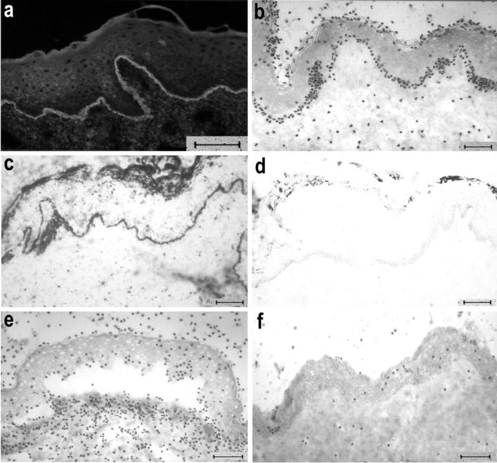
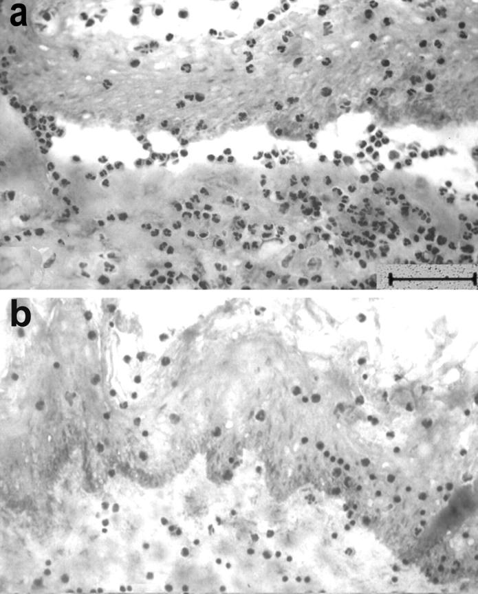
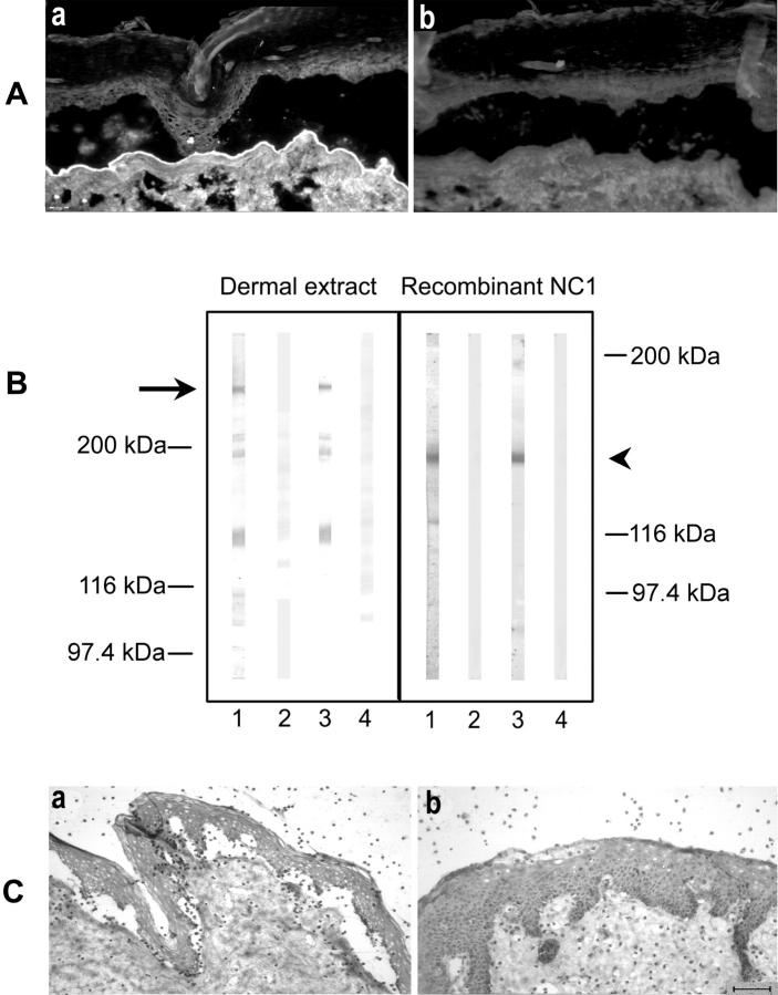
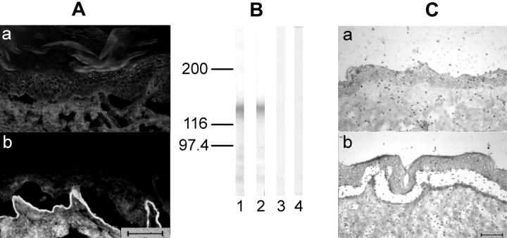
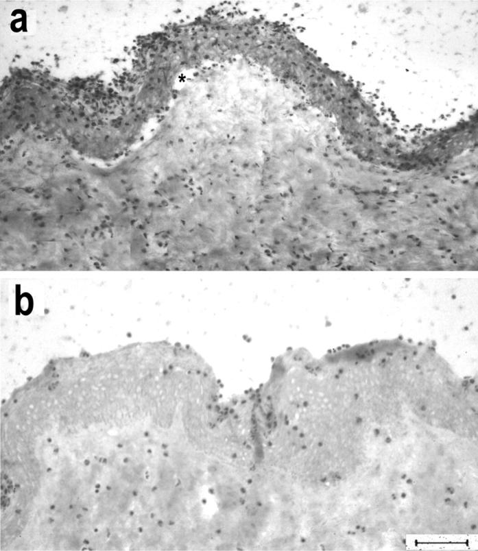
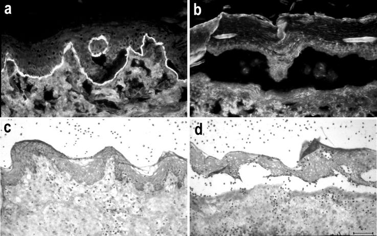
Epidermolysis bullosa acquisita is an autoimmune subepidermal blistering disease associated with autoantibodies to type VII collagen, the major constituent of anchoring fibrils. Previous attempts to demonstrate the blister-inducing potential of autoantibodies to this protein have failed. To address this question, we used an in vitro model involving cryosections of human skin incubated with patients' autoantibodies and leukocytes from healthy donors. We show that sera from 14 of 16 epidermolysis bullosa acquisita patients, in contrast to sera from healthy controls, induced dermal-epidermal separation in the cryosections. Recruitment and activation of neutrophils at the dermal-epidermal junction was necessary for split induction, whereas mononuclear cells were not required. Importantly, patients' autoantibodies affinity-purified against a recombinant form of the noncollagenous 1 domain of type VII collagen retained their blister-inducing capacity in a dose-dependent manner, whereas patients' IgG that was depleted of reactivity to type VII collagen lost this ability. Monoclonal antibody LH7.2 to the noncollagenous 1 domain of type VII collagen also induced subepidermal splits in the cryosections; F(ab')(2) fragments of autoantibodies to type VII collagen were not pathogenic. We demonstrate the capacity of autoantibodies to type VII collagen to trigger an Fcgamma-dependent inflammation leading to split formation in cryosections of human skin.


Figures

Figure 1.
Figure 1.
Serum from an EBA patient recruits neutrophils to the DEJ and induces subepidermal splits. a: Cryosections of normal human skin, incubated with EBA serum, show IgG deposits at the DEJ by indirect IF microscopy. b: Subsequent addition of leukocytes leads to neutrophil recruitment at the DEJ. Activation of neutrophils, as revealed by reduction of nitro blue tetrazolium to formazan (dark precipitates), is induced by EBA serum (c), but not by normal human serum (d). e: With longer incubation times, subepidermal splits develop in the cryosections that were treated with EBA serum. f: In contrast, serum from a healthy donor does not induce neutrophil recruitment or dermal-epidermal separation. Scale bars, 40 μm.
Figure 2.
Figure 2.
Granulocytes, but not mononuclear cells, are required for split induction by autoantibodies from EBA patients in cryosections of human skin. a: Granulocytes, which were isolated from the blood of healthy donors, mediate subepidermal cleavage in cryosections treated with an EBA serum. b: In contrast, mononuclear cells are not recruited to the DEJ and do not cause subepidermal cleavage.
Figure 3.
Figure 3.
Immunoadsorption of EBA serum against the recombinant NC1 domain of type VII collagen abolishes split induction. Autoantibodies to type VII collagen from EBA serum were purified using a recombinant form of the NC1 domain covalently coupled to an agarose matrix. A: When analyzed by indirect IF microscopy, IgG eluted from recombinant NC1 retains reactivity with the dermal side of NaCl-split human skin (a), in contrast to IgG from the flow-through fraction that completely lost IF reactivity (b). B: By immunoblotting, IgG autoantibodies eluted from the column (lane 3) recognized, like a serum sample from the same patient (lane 1), both full-length type VII collagen extracted from dermis (arrow) and a recombinant form of its NC1 domain (arrowhead). In contrast, the flow-through fraction (lane 2) or normal serum (lane 4) showed no reactivity. C: When incubated with the cryosections, the patient’s autoantibodies specific to type VII collagen NC-1 induce dermal-epidermal separation (a), whereas IgG depleted of reactivity to the NC1 domain (flow-through fraction) does not (b). Scale bar, 40 μm.
Figure 4.
Figure 4.
Preadsorption of EBA serum against proteins from insect cells infected with recombinant BV-0, lacking NC1-cDNA, does not abolish its blister-inducing capacity. EBA serum was purified against proteins from insect cells infected with recombinant BV-0 coupled to an agarose matrix. A: By indirect IF microscopy, IgG eluted from the column that completely lost indirect IF reactivity (a), in contrast IgG from the flow-through fraction stained the dermal side of salt-split skin (b). B: By immunoblotting, like the original serum (lane 1), the flow-through fraction (lane 2), reacted with recombinant NC1, whereas the eluted fraction (lane 3), like normal human serum (lane 4), was unreactive. C: When incubated with cryosections, in contrast to the eluted fraction (a), the flow-through fraction retained its ability to induce blisters (b). Scale bars, 40 μm.
Figure 5.
Figure 5.
A mAb to the NC1 domain of type VII collagen induces subepidermal splits in cryosections of human skin. Incubation of cryosections with mAb LH7.2, directed to the NC1 domain, and subsequently with leukocytes leads to dermal-epidermal separation (a), in contrast to a mAb to type IV collagen that was used at the same IgG concentration (b). Asterisk indicates the cleavage. Scale bar, 40 μm.
Figure 6.
Figure 6.
Dermal-epidermal separation is dependent on the Fc portion of autoantibodies to type VII collagen. Patient’s autoantibodies, which had been purified against recombinant NC1 of type VII collagen, were subsequently subjected to pepsin digestion. Resulting F(ab′)2 fragments labeled the dermal side of 1 mol/L of NaCl-split skin by indirect IF microscopy using an anti-Fab fluorescein isothiocyanate-conjugated secondary antibody (a), whereas no staining was observed using a Fc-specific conjugate (b). F(ab′)2 fragments failed to induce blisters (c), in contrast to unfragmented autoantibodies to type VII collagen (d). Scale bar, 40 μm.

References

    1. Woodley DT, Gammon WR, Briggaman RA: Epidermolysis bullosa acquisita 1999. Freedberg IM Eisen AZ Wolff K Austen KF Goldsmith LA Katz SI Fitzpatrick TB eds. Fitzpatrick’s Dermatology in General Medicine, ed 4 1999:pp 702-709 McGraw-Hill, New York
    1. Gammon WR, Briggaman RA, Inman AOD, Queen LL, Wheeler CE: Differentiating anti-lamina lucida and anti-sublamina densa anti-BMZ antibodies by indirect immunofluorescence on 1.0 M sodium chloride-separated skin. J Invest Dermatol 1984, 82:139-144 - PubMed
    1. Yaoita H, Briggaman RA, Lawley TJ, Provost TT, Katz SI: Epidermolysis bullosa acquisita: ultrastructural and immunological studies. J Invest Dermatol 1981, 76:288-292 - PubMed
    1. Woodley DT, Briggaman RA, O’Keefe EJ, Inman AO, Queen LL, Gammon WR: Identification of the skin basement-membrane autoantigen in epidermolysis bullosa acquisita. N Engl J Med 1984, 310:1007-1013 - PubMed
    1. Shimizu H, Ishiko A, Masunaga T, Kurihara Y, Sato M, Bruckner-Tuderman L, Nishikawa T: Most anchoring fibrils in human skin originate and terminate in the lamina densa. Lab Invest 1997, 76:753-763 - PubMed

Publication types

MeSH terms

LinkOut - more resources