. 2002 Aug;184(15):4277-87.
doi: 10.1128/JB.184.15.4277-4287.2002.

Functional characterization of Gne (UDP-N-acetylglucosamine-4-epimerase), Wzz (chain length determinant), and Wzy (O-antigen polymerase) of Yersinia enterocolitica serotype O:8

Affiliations

Functional characterization of Gne (UDP-N-acetylglucosamine-4-epimerase), Wzz (chain length determinant), and Wzy (O-antigen polymerase) of Yersinia enterocolitica serotype O:8

José Antonio Bengoechea et al. J Bacteriol. 2002 Aug.

Abstract

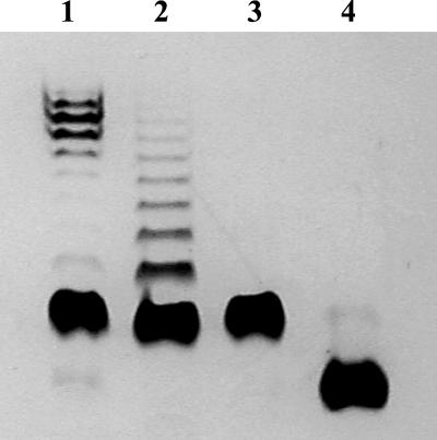
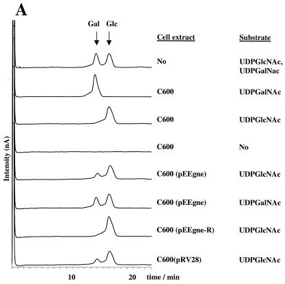
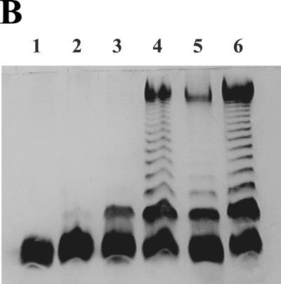
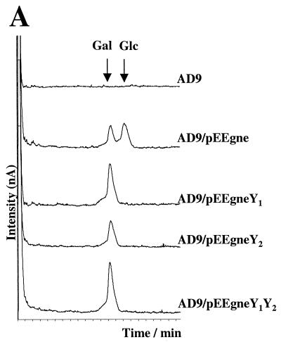
The lipopolysaccharide (LPS) O-antigen of Yersinia enterocolitica serotype O:8 is formed by branched pentasaccharide repeat units that contain N-acetylgalactosamine (GalNAc), L-fucose (Fuc), D-galactose (Gal), D-mannose (Man), and 6-deoxy-D-gulose (6d-Gul). Its biosynthesis requires at least enzymes for the synthesis of each nucleoside diphosphate-activated sugar precursor; five glycosyltransferases, one for each sugar residue; a flippase (Wzx); and an O-antigen polymerase (Wzy). As this LPS shows a characteristic preferred O-antigen chain length, the presence of a chain length determinant protein (Wzz) is also expected. By targeted mutagenesis, we identify within the O-antigen gene cluster the genes encoding Wzy and Wzz. We also present genetic and biochemical evidence showing that the gene previously called galE encodes a UDP-N-acetylglucosamine-4-epimerase (EC 5.1.3.7) required for the biosynthesis of the first sugar of the O-unit. Accordingly, the gene was renamed gne. Gne also has some UDP-glucose-4-epimerase (EC 5.1.3.2) activity, as it restores the core production of an Escherichia coli K-12 galE mutant. The three-dimensional structure of Gne was modeled based on the crystal structure of E. coli GalE. Detailed structural comparison of the active sites of Gne and GalE revealed that additional space is required to accommodate the N-acetyl group in Gne and that this space is occupied by two Tyr residues in GalE whereas the corresponding residues present in Gne are Leu136 and Cys297. The Gne Leu136Tyr and Cys297Tyr variants completely lost the UDP-N-acetylglucosamine-4-epimerase activity while retaining the ability to complement the LPS phenotype of the E. coli galE mutant. Finally, we report that Yersinia Wzx has relaxed specificity for the translocated oligosaccharide, contrary to Wzy, which is strictly specific for the O-unit to be polymerized.


Figures

FIG. 1.
FIG. 1.
DOC-PAGE analysis followed by silver staining of LPS preparations from Y. enterocolitica O:8 strains. Lane 1, strain YeO8c; lane 2, YeO8c-ΔWzzGB; lane 3, YeO8c-WbcEGB; lane 4, 8081-R1.
FIG. 2.
FIG. 2.
Analysis of UDP-N-acetylglucosamine-4-epimerase activity. (A) Dionex analysis of the UDP-N-acetylglucosamine-4-epimerase activities of different E. coli C600 cell extracts. The sources of the cell extracts and the substrates used in each reaction are shown at the right. The positions of the UDP-GalNAc (Gal) and UDP-GlcNAc (Glc) peaks are indicated by vertical arrows. (B) DOC-PAGE analysis followed by silver staining of LPS preparations from different E. coli strains. Lane 1, C600; lane 2, C600(pLZ6010); lane 3, C600(pLZ6010, pEEgne-R); lane 4, C600(pLZ6010, pRV28); lane 5, C600(pLZ6010, pEEgne); lane 6, C600(pLZ6020). The apparent difference between samples run in lanes 2 and 3 is due to an unequal loading of the sample run in lane 2.
FIG. 2.
FIG. 2.
Analysis of UDP-N-acetylglucosamine-4-epimerase activity. (A) Dionex analysis of the UDP-N-acetylglucosamine-4-epimerase activities of different E. coli C600 cell extracts. The sources of the cell extracts and the substrates used in each reaction are shown at the right. The positions of the UDP-GalNAc (Gal) and UDP-GlcNAc (Glc) peaks are indicated by vertical arrows. (B) DOC-PAGE analysis followed by silver staining of LPS preparations from different E. coli strains. Lane 1, C600; lane 2, C600(pLZ6010); lane 3, C600(pLZ6010, pEEgne-R); lane 4, C600(pLZ6010, pRV28); lane 5, C600(pLZ6010, pEEgne); lane 6, C600(pLZ6020). The apparent difference between samples run in lanes 2 and 3 is due to an unequal loading of the sample run in lane 2.
FIG. 3.
FIG. 3.
Molecular modeling of the active sites of the Gne and GalE type 4 epimerases. (A) Superimposition of the Y. enterocolitica Gne active site with UDP-glucose (yellow; the 2′-hydroxyl group is cyan) and UDP-galactose (green; the 2′-hydroxyl group is white). The residues involved in sugar binding are shown as ball-and-stick diagrams. (B) Superimposition of the UDP-galactose (carbon atoms are green) complexes of Y. enterocolitica Gne and E. coli GalE. The conserved sugar binding residues are shown as ball-and-stick diagrams. The residues Cys297 and Leu136 in Y. enterocolitica Gne (cyan) are replaced with tyrosines (magenta) in E. coli GalE. The 2′-hydroxyl group of UDP-galactose is white. (C) Empty volume (cyan) in the active site of the Y. enterocolitica Gne/NADH/UDP-galactose model. The cavity is large enough to accommodate the aminoacetyl group of UDP-GalNAc. The residues Cys297 and Leu136 in Y. enterocolitica Gne (cyan) are replaced with tyrosines (magenta) in E. coli GalE.
FIG. 4.
FIG. 4.
Critical role of the Cys297 and Leu136 residues in UDP-N-acetylglucosamine-4-epimerase activity of Gne. (A) Dionex analysis of the UDP-N-acetylglucosamine-4-epimerase activities of different cell extracts. The sources of the cell extracts are shown at the right. The amino acid changes introduced in the Gne variants are Leu136Tyr in pEEgneY1, Cys297Tyr in pEEgneY2, and both in pEEgneY1Y2. UDP-GalNAc was used as a substrate in all reactions. The positions of the UDP-GalNAc (Gal) and UDP-GlcNAc (Glc) peaks are indicated by vertical arrows. (B) Sodium dodecyl sulfate-PAGE analysis followed by silver staining of LPS preparations from different E. coli strains. Lane 1, E. coli AD9(pEEgneY1Y2); lane 2, E. coli AD9(pEEgneY2); lane 3, E. coli AD9(pEEgneY1); lane 4, E. coli AD9(pEEgne); lane 5, E. coli AD9; lane 6, E. coli C600.
FIG. 4.
FIG. 4.
Critical role of the Cys297 and Leu136 residues in UDP-N-acetylglucosamine-4-epimerase activity of Gne. (A) Dionex analysis of the UDP-N-acetylglucosamine-4-epimerase activities of different cell extracts. The sources of the cell extracts are shown at the right. The amino acid changes introduced in the Gne variants are Leu136Tyr in pEEgneY1, Cys297Tyr in pEEgneY2, and both in pEEgneY1Y2. UDP-GalNAc was used as a substrate in all reactions. The positions of the UDP-GalNAc (Gal) and UDP-GlcNAc (Glc) peaks are indicated by vertical arrows. (B) Sodium dodecyl sulfate-PAGE analysis followed by silver staining of LPS preparations from different E. coli strains. Lane 1, E. coli AD9(pEEgneY1Y2); lane 2, E. coli AD9(pEEgneY2); lane 3, E. coli AD9(pEEgneY1); lane 4, E. coli AD9(pEEgne); lane 5, E. coli AD9; lane 6, E. coli C600.
FIG. 5.
FIG. 5.
DOC-PAGE analysis followed by silver staining of LPS preparations from different E. coli strains. Lane 1, E. coli Sφ874(pLZ6020); lane 2, E. coli Sφ874(pLZ6010); lane 3, E. coli Sφ874; lane 4, E. coli C600(pLZ6010); lane 5, E. coli C600.

References

    1. Alexander, D. C., and M. A. Valvano. 1994. Role of the rfe gene in the biosynthesis of the Escherichia coli O7-specific lipopolysaccharide and other O-specific polysaccharides containing N-acetylglucosamine. J. Bacteriol. 176:7079-7084. - PMC - PubMed
    1. Appleyard, R. K. 1954. Segregation of new lysogenic types during growth of doubly lysogenic strain derived from Escherichia coli K12. Genetics 39:440-452. - PMC - PubMed
    1. Bastin, D. A., G. Stevenson, P. K. Brown, A. Haase, and P. R. Reeves. 1993. Repeat unit polysaccharides of bacteria—a model for polymerization resembling that of ribosomes and fatty acid synthetase, with a novel mechanism for determining chain length. Mol. Microbiol. 7:725-734. - PubMed
    1. Batchelor, R. A., P. Alifano, E. Biffali, S. I. Hull, and R. A. Hull. 1992. Nucleotide sequences of the genes regulating O-polysaccharide antigen chain length (rol) from Escherichia coli and Salmonella typhimurium—protein homology and functional complementation. J. Bacteriol. 174:5228-5236. - PMC - PubMed
    1. Batchelor, R. A., G. E. Haraguchi, R. A. Hull, and S. I. Hull. 1991. Regulation by a novel protein of the bimodal distribution of lipopolysaccharide in the outer membrane of Escherichia coli. J. Bacteriol. 173:5699-5704. - PMC - PubMed

Publication types

MeSH terms