Comparative Study
. 2002 Jul 23;99(15):9679-84.
doi: 10.1073/pnas.142310499. Epub 2002 Jul 9.

Genomic evidence that the intracellular proteins of archaeal microbes contain disulfide bonds

Affiliations
Comparative Study

Genomic evidence that the intracellular proteins of archaeal microbes contain disulfide bonds

Parag Mallick et al. Proc Natl Acad Sci U S A. .

Abstract

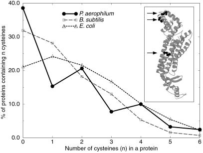
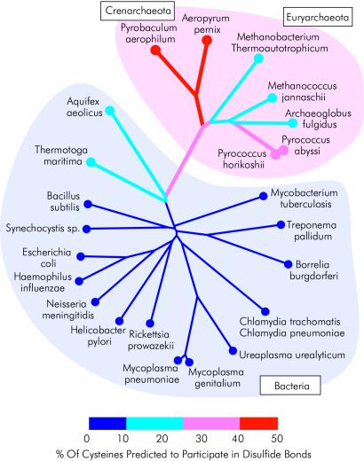
Disulfide bonds have only rarely been found in intracellular proteins. That pattern is consistent with the chemically reducing environment inside the cells of well-studied organisms. However, recent experiments and new calculations based on genomic data of archaea provide striking contradictions to this pattern. Our results indicate that the intracellular proteins of certain hyperthermophilic archaea, especially the crenarchaea Pyrobaculum aerophilum and Aeropyrum pernix, are rich in disulfide bonds. This finding implicates disulfide bonding in stabilizing many thermostable proteins and points to novel chemical environments inside these microbes. These unexpected results illustrate the wealth of biochemical insights available from the growing reservoir of genomic data.


Figures

Figure 1
Figure 1
An abundance of intracellular disulfide bonds in P. aerophilum is suggested by the preference for even numbers of cysteine residues in its proteins (bold line). For controls, corresponding plots are shown for B. subtilis (dashed line) and E. coli (dotted line). In this plot, data are drawn from proteins ranging in size from 100 to 200 amino acids, as the phenomenon is most evident for small to medium-ized proteins. Inset shows the structure of adenylosuccinate lyase from P. aerophilum (12). Its three disulfide bonds led to the initial suggestion that some hyperthermophilic archaea might be rich in intracellular protein disulfide bonds.
Figure 2
Figure 2
A sequence-structure mapping method for estimating disulfide abundance in a set of protein sequences. For each protein sequence in a genome, a homologous structure is identified if one is available in the Protein Data Bank, by using blast, PSI-BLAST and the Method of Sequence Derived Properties (22, 23). Each sequence is mapped onto its homologous three-dimensional protein structure. The spatial proximity of each pair of cysteines is then examined to determine whether the two could potentially form a disulfide bond. The total disulfide abundance for a genome is estimated statistically by comparison with results on sets of control proteins as described in Materials and Methods.
Figure 3
Figure 3
The Triangle Method for searching for possible disulfide bonds in proteins from P. aerophilum. Triplets of similar proteins are found by blast, one of which is from P. aerophilum, a second from E. coli, and the third of known structure. Here the protein of known structure is PCMT from Thermotoga maritima (Lower Center). The three sequences align convincingly (fragment of alignment shown at the top). Two additional cysteine residues present in the P. aerophilum sequence are indicated by boxes. The model on the upper left shows that the P. aerophilum sequence, when mapped on the T. maritima structure, places these two cysteine residues (circled, shown in black) close enough to form a disulfide bond. In the E. coli model (Upper Right), neither of the corresponding residues is cysteine, and no disulfide bond can form here or anywhere else in the E. coli enzyme.
Figure 4
Figure 4
A fluorescent image of an SDS/PAGE gel showing abundant disulfide bonds in the proteins of P. aerophilum. Cysteine residues in their free sulfhydryl form were selectively labeled with the fluorescent reagent, CPM, either in the absence of (lanes 1 and 3) or after (lanes 2 and 4) reduction of disulfides with TCEP. E. coli was chosen as a control organism and shows very few protein disulfide bonds, as expected (lanes 1 and 2). The negative of the image is shown for improved visualization of labeled bands.
Figure 5
Figure 5
An unrooted phylogenetic tree showing the 25 genomes whose disulfide bond abundances were estimated computationally. The thickness and color of each branch illustrate the predicted disulfide content of the microbe at the branch's terminus according to the key (bottom); the coloring of internal branches reflects an average over multiple species. Archaeal organisms lie within the pink region. Bacterial genomes lie within the light blue region. The crenarchaeal branches of the tree (deep red, thickest) have the greatest predicted disulfide content, exceeding 40%. The majority of bacteria have low predicted disulfide contents ranging from 0 to 10% (deep blue, thin lines). The thermophilic eubacteria, Aquifex and Thermotoga, have predicted disulfide contents in the 10–20% range, whereas the euryarchaea have values ranging from 10 to 30%. Other methods of construction can lead to phylogenetic trees that differ in detail from the one shown here without affecting the salient features.

References

    1. Marcotte E M, Pellegrini M, Ng H L, Rice D W, Yeates T O, Eisenberg D. Science. 1999;285:751–753. - PubMed
    1. Lin J, Gerstein M. Genome Res. 2000;10:808–818. - PMC - PubMed
    1. Fraser C M, Eisen J, Fleischmann R D, Ketchum K A, Peterson S. Emerg Infect Dis. 2000;6:505–512. - PMC - PubMed
    1. Sunyaev S, Lathe W, Bork P. Curr Opin Struct Biol. 2001;11:125–130. - PubMed
    1. Wolf Y I, Brenner S E, Bash P A, Koonin E V. Genome Res. 1999;9:17–26. - PubMed

Publication types

LinkOut - more resources