. 2002 Jul 23;99(15):9990-5.
doi: 10.1073/pnas.152706299. Epub 2002 Jul 9.

A constitutively active dioxin/aryl hydrocarbon receptor induces stomach tumors

Affiliations

A constitutively active dioxin/aryl hydrocarbon receptor induces stomach tumors

Patrik Andersson et al. Proc Natl Acad Sci U S A. .

Abstract

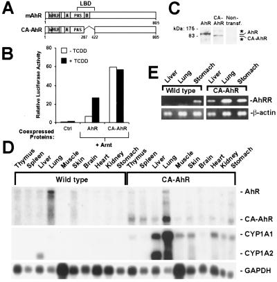
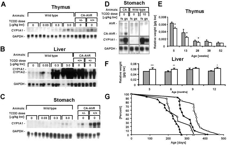
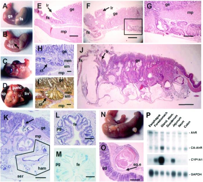
The dioxin/aryl hydrocarbon receptor (AhR) functions as a ligand-activated transcription factor regulating transcription of a battery of genes encoding xenobiotic metabolizing enzymes. Known receptor ligands are environmental pollutants including polycyclic aromatic hydrocarbons and polychlorinated dioxins. Loss-of-function (gene-disruption) studies in mice have demonstrated that the AhR is involved in toxic effects of dioxins but have not yielded unequivocal results concerning the physiological function of the receptor. Gain-of-function studies therefore were performed to unravel the biological functions of the AhR. A constitutively active AhR expressed in transgenic mice reduced the life span of the mice and induced tumors in the glandular part of the stomach, demonstrating the oncogenic potential of the AhR and implicating the receptor in regulation of cell proliferation.


Figures

Figure 1
Figure 1
Constitutive activity of CA-AhR. (A) Schematic representation of the wild-type mouse AhR (mAhR) and of CA-AhR. bHLH, basic helix–loop–helix; PAS, Per-Arnt-Sim. (B) Functional activity of CA-AhR in Chinese hamster ovary cells. Cells were transiently transfected with an AhR-dependent luciferase reporter gene and expression vectors encoding AhR nuclear translocator (Arnt), wild-type AhR, or CA-AhR. The control lanes (Ctrl) represent activity from the reporter gene alone and empty expression vector. Data are from one experiment performed in duplicate and are representative of at least three independent experiments. (C) Detection of the AhR and CA-AhR proteins expressed after transient transfection of Chinese hamster ovary cells. Whole-cell extracts were analyzed by immunoblotting using anti-AhR antibodies. The star indicates nonspecific immunoreactivity. (D) Expression and functional activity of CA-AhR in 8-month-old female mice: RNA blot analysis showing expression of the endogenous AhR, CA-AhR, and the target genes CYP1A1 and CYP1A2. The expression of glyceraldehyde-3-phosphate dehydrogenase (GAPDH) is shown as RNA-loading control of corresponding tissues. (E) AhR repressor (AhRR) and β-actin mRNA expression was assessed by reverse transcription–PCR.
Figure 2
Figure 2
Functional activity of CA-AhR. (AC) RNA blots showing expression of CYP1A1 and glyceraldehyde-3-phosphate dehydrogenase (GAPDH) mRNA in thymus, liver, and stomach of 6-month-old female wild-type, heterozygous, and homozygous CA-AhR mice treated orally 3 days earlier with vehicle or TCDD as indicated. bw, body weight. (D) RNA blot showing expression of AhR, CA-AhR, CYP1A1, and glyceraldehyde-3-phosphate dehydrogenase in forestomach (fs) or glandular stomach (gs) from 3-month-old CA-AhR (CA) or wild-type male mice treated orally with vehicle or TCDD 1 day earlier. (E and F) Alteration of the relative weights of thymus and liver in homozygous CA-AhR animals. The closed bars represent wild-type animals, and open bars represent CA-AhR animals. Thymuses from at least four female animals of each genotype and age and livers from at least five males of each genotype and age were examined. *, P < 0.05, **, P < 0.01, and ***, P < 0.005 as assessed by two-tailed Student's t test. (G) Ages of the homozygous CA-AhR mice found dead stratified by sex (closed symbols for males, open for females) and independent founder lines (triangles for line A3 and circles for line Y8).
Figure 3
Figure 3
Neoplastic lesions and intestinal metaplasia in the stomach of CA-AhR mice. (A) Normal stomach from a 12-month-old wild-type male showing the forestomach (fs) and glandular stomach (gs). (B) At 3–4 months of age single small cysts close to the limiting ridge were seen in CA-AhR mice (arrow). (C) In older CA-AhR animals (6–12 months old) the number of cystic tumors increased and occupied a larger area of the stomach. (D) In the most severe cases (9–12 months of age), the stomach was adherent to adjacent organs such as spleen (sp), pancreas (panc), and liver (liv). (E) Normal stomach from a 6-month-old wild-type male mouse showing the muscularis propria layer (mp) and limiting ridge (lr) constituting the border between the squamous epithelium of the forestomach and the glandular epithelium (ge, HE staining). (Bar, 0.5 mm.) (F) Close to the limiting ridge a rupture of the submucosa by neoplastic crypts is seen in a 3.5-month-old CA-AhR male. Note glands within the stroma of the limiting ridge (HE). (Bar, 0.5 mm.) (G) Larger magnification of the boxed area in F (HE). (Bar, 0.15 mm.) (H and I) Invading crypts surrounded by connective tissue (ct) invade the submucosa (sm) by penetrating through the muscularis mucosa (mm) and submucosa layers and further into the muscularis propria in a 9-month-old CA-AhR female (H, HE staining; I, van Gieson staining). (Bars, 0.1 mm.) (J) Stomach from a 12-month-old CA-AhR male with severely distorted tissue architecture (HE). (Bar, 1.25 mm.) (K) Glands underlying the serosa (ser) in a 12-month-old CA-AhR female with characteristics of a hamartoma (ham), i.e., lymphatic tissue, vessels, and fat (HE). (Bar, 0.5 mm.) Note also invasion (arrow) of glands from the glandular epithelium into the muscularis propria. (L and M) Glandular structures located in the muscularis propria with cells resembling foveolar epithelium (fe) and pyloric glands (pg) showing intestinal metaplasia in a 9-month-old CA-AhR male (L, HE staining; M, Alcian blue pH 2.5 staining). (Bars, 0.1 mm.) (N and O) Squamous cysts on the caecum showing colonic glands (cg) and squamous epithelium (sq.e) of a 9-month-old CA-AhR male (HE). (Bar, 0.5 mm.) (P) The expression and activity of CA-AhR in the gastrointestinal tract is highest in the glandular stomach. RNA blot showing expression of CA-AhR, endogenous AhR (AhR), CYP1A1, and glyceraldehyde-3-phosphate dehydrogenase (GAPDH) mRNA in different parts of the alimentary tract of homozygous CA-AhR mice 3 months of age.
Figure 4
Figure 4
Staining for proliferation, target-gene induction, and AhR expression in glandular stomachs from control or TCDD-treated wild-type mice and CA-AhR mice. Nine-month-old wild-type (wt) or CA-AhR males were treated orally with vehicle (oil) or 10 μg of TCDD/kg of body weight, and tissues were removed and fixed in formaldehyde 4 days later. Paraffin-embedded serial sections were stained with HE (AC) or immunostained with antibodies specific for PCNA (DF), CYP1A1 (GI), or AhR (JO). Incubation without primary antibody was used as negative control. (Bars in AL, 0.25 mm; bars in MO, 0.015 mm.) The brackets, arrows, and star indicate reference points in the serial sections. (DF) The proliferative zone shown by PCNA staining (brackets) is expanded basally into the parietal/chief cell region in stomachs from TCDD-treated wild-type animals and CA-AhR animals. The arrow indicates proliferation in submucosal tumors in the CA-AhR stomach. (GI) Examples of CYP1A1 staining are shown by arrows. The star indicates strong CYP1A1 expression in the submucosal tumor. (JO) AhR is expressed throughout the glandular stomach of both wild-type and CA-AhR mice. The arrow in L indicates a submucosal tumor. The arrow in O indicates mainly nuclear staining in the stomach of CA-AhR mice.
Figure 5
Figure 5
Severity of the tumors correlated with CA-AhR gene dosage and sex. Tissue sections were used to classify in arbitrary units the cellular alterations from 0 (for no cystic/dysplastic histological changes) to 5 (for severe cystic/dysplastic histological changes). (A) The histological classification of stomach tumors in 12-month-old wild-type, heterozygous (+/−), and homozygous (+/+) CA-AhR male mice demonstrates a gene-dosage effect. The stomach lesions became more severe with increasing age of both sexes, with homozygous male mice (B) being more susceptible than homozygous female mice (C).

References

    1. Gu Y Z, Hogenesch J B, Bradfield C A. Annu Rev Pharmacol Toxicol. 2000;40:519–561. - PubMed
    1. Pohjanvirta R, Tuomisto J. Pharmacol Rev. 1994;46:483–549. - PubMed
    1. Shimizu Y, Nakatsuru Y, Ichinose M, Takahashi Y, Kume H, Mimura J, Fujii-Kuriyama Y, Ishikawa T. Proc Natl Acad Sci USA. 2000;97:779–782. - PMC - PubMed
    1. Fernandez-Salguero P, Pineau T, Hilbert D M, McPhail T, Lee S S, Kimura S, Nebert D W, Rudikoff S, Ward J M, Gonzalez F J. Science. 1995;268:722–726. - PubMed
    1. Schmidt J V, Su G H, Reddy J K, Simon M C, Bradfield C A. Proc Natl Acad Sci USA. 1996;93:6731–6736. - PMC - PubMed

Publication types

MeSH terms