. 2002 Jul 15;21(14):3632-42.
doi: 10.1093/emboj/cdf382.

Ferrichrome induces endosome to plasma membrane cycling of the ferrichrome transporter, Arn1p, in Saccharomyces cerevisiae

Affiliations

Ferrichrome induces endosome to plasma membrane cycling of the ferrichrome transporter, Arn1p, in Saccharomyces cerevisiae

Youngwoo Kim et al. EMBO J. .

Abstract

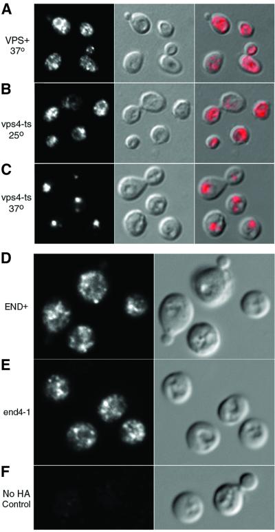
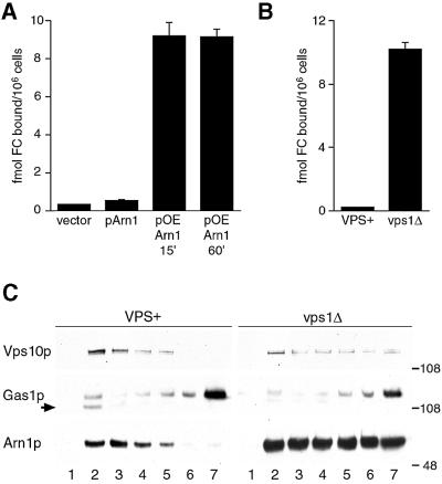
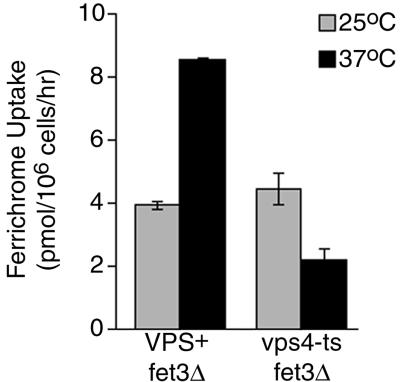
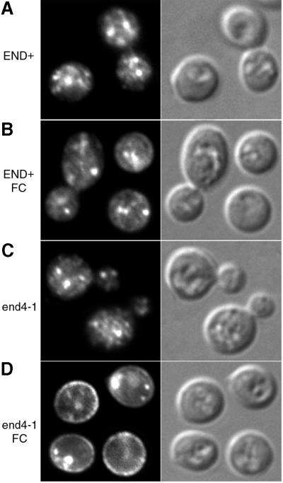
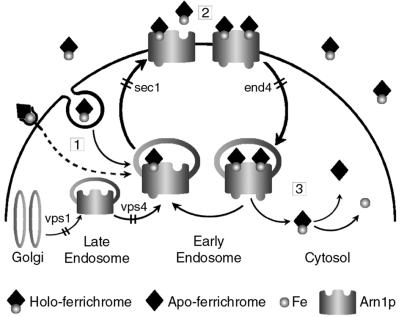
Siderophores are small iron-binding molecules that are synthesized and secreted in the iron-free form by microorganisms. Saccharomyces cerevisiae takes up iron bound to siderophores by two separate systems, one of which requires the ARN family of sidero phore-iron transporters. Arn1p and Arn3p are expressed in endosome-like intracellular vesicles. Here we present evidence that, in the absence of its specific substrate, ferrichrome, Arn1p is sorted directly from the Golgi to the endosomal compartment and does not cycle to the plasma membrane. When cells are exposed to ferrichrome at low concentrations, Arn1p stably relocalizes to the plasma membrane. At higher concentrations of ferrichrome, Arn1p relocalizes to the plasma membrane and rapidly undergoes endocytosis. Plasma membrane localization of Arn1p occurs only in the presence of its specific substrate, and not in the presence of other siderophores. Despite expression of Arn1p on the plasma membrane, mutant strains with defects in endocytosis exhibit reduced uptake of ferrichrome-iron. Thus, siderophores influence the trafficking of the Arn transporters within the cell and this trafficking is important for transporter function.


Figures

None
Fig. 1. Mislocalization of Arn1p in a vps4-ts strain and localization to the endosome in END+ and end4-1 strains. (AC) Strains SEY6210 (VPS+) and MBY3 (vps4-ts) were transformed with pArn1-HA and grown in iron-poor medium at 25°C. Aliquots of culture were shifted to 37°C (A and C) for 1 h or grown at 25°C (B) prior to fixation and preparation for indirect immunofluorescence. Mouse monoclonal HA.11 was the primary antibody and Cy3-conjugated donkey anti-mouse was the secondary antibody. Images are in sets of three: fluorescence on the left, differential interference contrast (DIC) in the center and the merged image on the right. (D and E) Congenic RH144–3D (END+; D) and RH268–1C (end4-1; E) strains were transformed with pMetArn1-HA and grown in iron-poor medium at 22°C. Cells were shifted to methionine-free, iron-poor medium and cultured at 37°C for 2 h prior to fixation and preparation for indirect immunofluorescence microscopy. A wild-type strain that did not carry an HA-tagged allele was treated identically, as a control (F). Images are in pairs with fluorescence on the left and DIC on the right.
None
Fig. 2. Mislocalization of Arn1p to the plasma membrane in a vps1Δ strain. (A and B) Binding of ferrichrome to Arn1p expressed on the plasma membrane. (A) Strain CWY101, in which all four Arn transporters have been deleted, was transformed with the vector pRS316 (vector), the low-copy-number pArn1-HA (pArn1) to reconstitute endogenous levels of Arn1p, or the high-copy-number pOE-Arn1-HA (pOE Arn1). Cells were grown to mid-log phase in iron-poor medium, then membrane transport and uptake systems were inactivated by incubation with NaN3 and KF at 0°C. The capacity of Arn1p that is expressed on the plasma membrane to bind [55Fe]ferrichrome at 0°C was measured. (B) Strain RH144–3D (VPS+) and the congenic strain CWY102 (vps1Δ) were grown to mid-log phase in rich medium and cell surface binding of [55Fe]ferrichrome was measured. (C) Expression of Arn1p in both plasma membrane and intracellular vesicle fractions in a vps1 deletion strain. Strain RH144-3D (VPS+) and the congenic strain CWY102 (vps1Δ) were transformed with pArn1-HA and grown to mid-log phase in rich medium. Cells were lysed, membranes were collected and then separated on discontinuous sucrose gradients. Fractions were collected from the top and subjected to SDS–PAGE and western blotting using antibodies directed against Vps10p to detect late-Golgi membranes (Vps10p), Gas1p to detect plasma membrane (Gas1p) and the HA epitope to detect Arn1p-HA (Arn1p). The arrow indicates an endoplasmic reticulum-derived, immature form of Gas1p. Molecular weight standards are indicated in kDa.
None
Fig. 3. Loss of the uptake of ferrichrome–iron in the vps4-ts strain. Congenic strains CWY106 (VPS+ fet3Δ) and CWY105 (vps4-ts fet3Δ) were grown to mid-log phase in rich media (YPD). Strains were either grown continuously at 25°C or shifted to 37°C for 1 h prior to the measurement of uptake of [55Fe]ferrichrome at the indicated temperatures, as described in Materials and methods. Experiments were repeated twice; data from a representative experiment are shown.
None
Fig. 4. Plasma membrane localization of Arn1p in the end4-1 strain in the presence of ferrichrome. Strains RH144-3D (END+) and RH268-1C (end4-1) were transformed with pMetArn1-HA and grown in iron-poor medium at 22°C. Cells were shifted to methionine-free, iron-poor medium at 37°C without (A and C) or with (B and D) 10 µM ferrichrome (FC) and grown for two additional hours prior to preparation for immunofluorescence microscopy. Images are in pairs with fluorescence on the left and DIC on the right.
None
Fig. 5. Concentration dependence of ferrichrome-induced plasma membrane localization of Arn1p. Parent strain RH144-3D carrying pMetArn1-HA (AE) and SEY5017 (sec1-1) carrying pArn1-HA (F) were grown in iron-poor medium at 22°C to mid-log phase. Cells were then transferred to methionine-free, iron-poor medium and exposed to ferrichrome at the indicated concentrations for 2 h prior to fixation and preparation for immunofluorescence microscopy. In (F), the sec1-1 strain was shifted to 37°C and exposed to 0.1 µM ferrichrome for 2 h prior to fixation and preparation for immunofluorescence microscopy. The identically treated parent strain (SEY6210, congenic to sec1-1) exhibited a fluorescence pattern identical to that shown in (C).
None
Fig. 6. Detection of Arn1p in the plasma membrane after exposure to ferrichrome. (A and B) Redistribution of Arn1p to fractions containing plasma membranes after exposure to ferrichrome. Strains RH144-3D (END+) and RH268-1C (end4-1) carrying pArn1-HA were grown in iron-poor medium at 22°C. Cells were then exposed to no ferrichrome, 0.1 µM or 10 µM ferrichrome and shifted to 37°C for two additional hours. Cells were lysed, membranes were collected and fractionated on sucrose step gradients, and the fractions subjected to SDS–PAGE and western blotting. In (A), lysates from cells not treated with ferrichrome were used and arrows indicate the distribution of late Golgi vesicles (Vps10p) and plasma membranes (Gas1p). In (B), the distribution of Arn1p was detected using anti-HA antibody. Protein molecular weight standards in kDa are indicated. (C) Binding of ferrichrome by Arn transporters localized to the plasma membrane. Strains RH144-3D (END+) and RH268-1C (end4-1) expressing only endogenous Arn transporters were grown and treated with ferrichrome as in (A) and (B). The binding of radiolabeled ferrichrome to the surface of the cells was measured as described in Materials and methods.
None
Fig. 7. Substrate-specific relocalization of Arn1p. Strains RH144-3D (END+; AF) and RH268-1C (end4-1; GL) were transformed with either pArn1-HA (A–C, G–I) or pArn3-HA (D–F, J–L) and grown at 22°C in iron-poor medium to mid-log phase. Cultures were shifted to 37°C, supplemented with no siderophore (No Siderophore; A, D, G and J), 10 µM ferrichrome (Ferrichrome; B, E, H and K) or 10 µM desferrioxamine B (Ferrioxamine B; C, F, I and L), and grown for an additional 2 h. Cells were then prepared for immunofluorescence microscopy. (M) Substrates of Arn1p and Arn3p. Siderophores taken up by Arn1p and Arn3p, as determined in siderophore–iron uptake assays, are indicated.
None
Fig. 8. Diminished uptake of ferrichrome–iron in yeast defective in endocytosis. (A–C) Strains of the indicated genotype were grown in rich medium (YPD) to mid-log phase. Cells were either grown continuously at 25°C or shifted to 37°C for 1 h prior to the measurement of ferrichrome–iron uptake (A and C) or ferrous iron uptake (B) at the indicated temperatures. (D) Strain CPY119 (fet3Δ) was grown in rich medium to mid-log phase and cells were treated with either 200 µM latrunculin-A (+LAT-A) or an equal volume of DMSO (–LAT-A) for 15 min prior to harvesting and measurement of ferrichrome–iron uptake. LAT-A or DMSO was also included in the uptake assay buffer.
None
Fig. 9. Proposed model for trafficking of Arn1p in the presence of ferrichrome. In step 1, the presence of extracellular ferrichrome is signaled to Arn1p as it cycles between the early and late endosome, which results in translocation of Arn1p to the plasma membrane. Ferrichrome may gain access to the endosome via fluid-phase endocytosis and directly bind Arn1p. Alternatively, receptor-mediated signaling events may indicate the presence of ferrichrome to the endosomal Arn1p. In step 2, Arn1p binds a second molecule of ferrichrome and subsequently undergoes endocytosis. In step 3, the ferrichrome–iron chelate is transported to the cytosol, where it undergoes reduction and release of iron. Binding of ferrichrome to each of the two sites may result in conformational changes in the transporter that affect sorting. Mutant strains used in this study are noted; the trafficking steps that are blocked by the mutations are indicated by a double bar.

Similar articles

Cited by

References

    1. Askwith C.C. and Kaplan,J. (1998) Site-directed mutagenesis of the yeast multicopper oxidase Fet3p. J. Biol. Chem., 273, 22415–22419. - PubMed
    1. Askwith C., Eide,D., Van Ho,A., Bernard,P.S., Li,L., Davis-Kaplan,S., Sipe,D.M. and Kaplan,J. (1994) The FET3 gene of S.cerevisiae encodes a multicopper oxidase required for ferrous iron uptake. Cell, 76, 403–410. - PubMed
    1. Ayscough K. (1998) Use of latrunculin-A, an actin monomer-binding drug. Methods Enzymol., 298, 18–25. - PubMed
    1. Babst M., Sato,T.K., Banta,L.M. and Emr,S.D. (1997) Endosomal transport function in yeast requires a novel AAA-type ATPase, Vps4p. EMBO J., 16, 1820–1831. - PMC - PubMed
    1. Bagnat M., Chang,A. and Simons,K. (2001) Plasma membrane proton ATPase pma1p requires raft association for surface delivery in yeast. Mol. Biol. Cell, 12, 4129–4138. - PMC - PubMed

MeSH terms