. 2002 Jul 15;21(14):3681-93.
doi: 10.1093/emboj/cdf372.

Selenoproteins and selenocysteine insertion system in the model plant cell system, Chlamydomonas reinhardtii

Affiliations

Selenoproteins and selenocysteine insertion system in the model plant cell system, Chlamydomonas reinhardtii

Sergey V Novoselov et al. EMBO J. .

Abstract

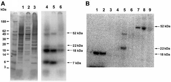
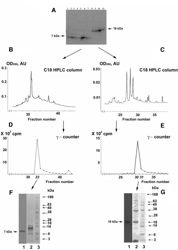
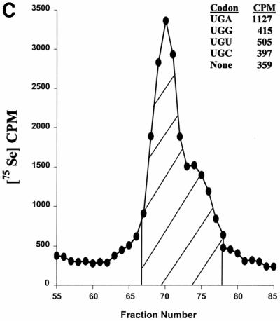
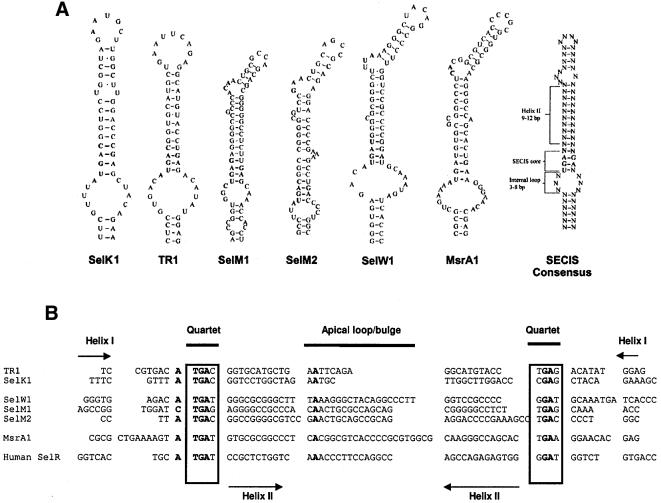
Known eukaryotic selenocysteine (Sec)-containing proteins are animal proteins, whereas selenoproteins have not been found in yeast and plants. Surprisingly, we detected selenoproteins in a member of the plant kingdom, Chlamydomonas reinhardtii, and directly identified two of them as phospholipid hydroperoxide glutathione peroxidase and selenoprotein W homologs. Moreover, a selenocysteyl-tRNA was isolated that recognized specifically the Sec codon UGA. Subsequent gene cloning and bioinformatics analyses identified eight additional selenoproteins, including methionine-S-sulfoxide reductase, a selenoprotein specific to Chlamydomonas: Chlamydomonas selenoprotein genes contained selenocysteine insertion sequence (SECIS) elements that were similar, but not identical, to those of animals. These SECIS elements could direct selenoprotein synthesis in mammalian cells, indicating a common origin of plant and animal Sec insertion systems. We found that selenium is required for optimal growth of Chlamydomonas: Finally, evolutionary analyses suggested that selenoproteins present in Chlamydomonas and animals evolved early, and were independently lost in land plants, yeast and some animals.


Figures

None
Fig. 1. Detection of Chlamydomonas selenoproteins. Chlamydomonas cells were grown in the presence of 75Se, collected by centrifugation, disrupted by sonication, and the resulting homogenate centrifuged at 18 000 g for 30 min. In (A), fractions were analyzed by SDS–PAGE. Lanes 1–3 (left panel) show homogenate, supernatant and pellet, respectively, stained with Coomassie Blue (protein markers are shown in the left-most lane and their sizes indicated on the left) and lanes 4–6 (right panel) show respective lanes in the same gel exposed to PhosphorImager detection of 75Se (sizes of detected selenoproteins are shown on the right). In (B), the soluble fraction [shown in (A), lane 2] was fractionated on a Q-Sepharose column. Lanes 1 and 2 show the flow-through fractions and contained the 7 and 18 kDa selenoproteins, and lanes 3–9 the fractions that eluted in the salt gradient (see Materials and methods). The 22 kDa selenoprotein shown in lane 5 eluted at ∼150 mM NaCl, and the 52 kDa selenoprotein shown in fractions 7–9 eluted at 400 mM NaCl in buffer B. Proteins were detected by PhosphorImager analysis of an SDS–PAGE gel.
None
Fig. 2. Purification of Chlamydomonas 7 and 18 kDa selenoproteins. The 7 and 18 kDa proteins were purified from the radioactive flow-through fractions from Q-Sepharose (see Figure 1B) by concentrating this material with ammonium sulfate, adjusting to a concentration of 0.4 M ammonium sulfate in buffer B, and fractionating on a phenyl–Sepharose HPLC column. Samples were analyzed as shown in (A): lanes 1–7 contain fractions eluted in a 0.4–0 M ammonium sulfate gradient in buffer B and lanes 8–11 show fractions eluted in a buffer B to water gradient as detected by PhosphorImager analysis; or further purified as shown in (B), (D) and (F): fractionation of the 7 kDa selenoprotein on a C18 reversed-phase HPLC column with a 0–70% acetonitrile gradient in 0.05% trifluoroacetic acid, γ-counter detection of 75Se in the eluted fraction, and SDS–PAGE analysis of fraction 33, respectively; and in (C), (E) and (G): fractionation of the 18 kDa selenoprotein on a C18 reversed-phase HPLC column with a 0–70% acetonitrile gradient in 0.05% trifluoroacetic acid, γ-counter detection of 75Se in the eluted fractions, and SDS–PAGE analysis of fraction 31, respectively. Lane 1 in (F) and (G) shows detection of 75Se by PhosphorImager analysis; lanes 2 and 3 show staining with Coomassie Blue of the column fractions and protein standards (masses are shown on the right).
None
Fig. 3. Selenoproteins and Sec tRNA in Chlamydomonas. (A) Alignment of human, Arabidopsis, Clostridium, C.elegans, Schistosoma mansoni, yeast and Chlamydomonas PHGPx. The sequences of five tryptic peptides of Chlamydomonas PHGPx1, for which amino acid sequences were determined experimentally, are underlined. The DDBJ/EMBL/GenBank accession Nos are: NP_002076.1, human PHGPx; BF936124.1, Schistosoma PHGPx; AE007667.1, Clostridium PHGPx; AE007667.1, S.cerevisiae PHGPx; NP_497078.1, C.elegans PHGPx; AV623602, Chlamydomonas PHGPx1; BI721156 and AV623602, Chlamydomonas PHGPx2. Sec is present in human, Schistosoma and Chlamydomonas proteins, but is replaced by Cys in other PHGPx homologs. (BChlamydomonas cDNA sequence encoding selenoprotein W1. The amino acid U represents selenocysteine-14, which is encoded by TGA (underlined). The sequences of three tryptic peptides, for which amino acid sequences were determined experimentally, are also underlined. In the 3′-UTR, the position of the SECIS element is shown. (C) Chromatography of Chlamydomonas [75Se]selenocysteyl-tRNA. Chlamydomonas cells (2.5 g) were labeled with 75Se, and the labeled tRNA extracted and chromatographed on an RPC-5 column as described in Materials and methods. The large peak and trailing shoulder of 75Se-containing tRNA were pooled, as shown by the hatched area in the figure, prepared for ribosomal binding studies and the ribosomal binding studies carried out as described in Materials and methods. Binding to ribosomes in the absence of codon (designated None) or in the presence of codon is shown in the figure. Total CPM added to each reaction were 3200.
None
Fig. 4. SECIS elements in Chlamydomonas selenoprotein genes. (A) SECIS structures. SECIS element structures are shown for selenoproteins indicated below the structures. Proposed Chlamydomonas SECIS element consensus is shown on the right. (B) Alignment of the SECIS elements of Chlamydomonas selenoproteins genes. TR1 and SelK1 structures are Form 1 structures and the others are Form 2 structures. Human SelR SECIS element is aligned for comparison. Conserved nucleotides are shown in bold. Locations of helix 1, helix 2, quartet and apical loop are indicated. (C) Expression of Chlamydomonas SelK1 in mammalian cells. NIH 3T3 cells were transfected with a plasmid encoding GFP-chlamySelK (lane 1) or with GFP-C3 Vector (Clontech) as control (lane 2). Transfected cells were grown in the presence of [75Se]selenite and the resulting 75Se-labeled proteins were resolved by SDS–PAGE and visualized with a PhosphorImager. The location of GFP–SelK1 product is shown on the left. Locations and molecular weights of major selenoproteins, thioredoxin reductase 1 (TR1) and glutathione peroxidase 1 (GPx1), are also indicated. (D) Multiple alignment of Chlamydomonas MsrA1. The accession numbers are: NP_495540.1, C.elegans MsrA; P54149, bovine MsrA; NP_036463.1, human MsrA; NP_290851.1, E.coli MsrA; AAK83645, A.thaliana MsrA; 11342533, Fragaria ananassa MsrA; 7446683, Schizosaccharomyces pombe MsrA; AF494053, C.reinhardtii MsrA1.
None
Fig. 5. Growth of Chlamydomonas in selenium-deficient and -sufficient media. Cells were grown for 110 h in selenium-deficient TAP medium and in the presence of 50 nM selenite as described in Materials and methods. Initial cell counts were 103 cells/ml. Changes in cell numbers were determined with a hemacytometer, and each sample was analyzed three times. Lines were drawn according to a ‘sigmoidal fit’ functions of Origin, version 5.0 (OriginLab Corporation, Northampton, MA). Selenium-deficient (open circles) and selenium-suplemented (closed circles) cells were grown in the presence of 0.5 mM H2O2 (B), 0.75 mM methionine-S-sulfoxide (C) or 0.5 mM H2O2 and 0.75 mM methionine-S-sulfoxide (D) or in the absence of these compounds (A).
None
Fig. 6. Phylogenetic analyses of Chlamydomonas selenoproteins. Each selenoprotein is shown in an evolutionary tree as follows. (A) SelW tree. (B) SelT tree. (C) SelK tree. (D) MsrA tree. (E) PHGPx tree. (F) SelM tree. Accession numbers are given in the legends to Figures 3A, 4D and Supplementary figures. Selenium-containing proteins are indicated by asterisks.

Similar articles

Cited by

References

    1. Arner E.S., Sarioglu,H., Lottspeich,F., Holmgren,A. and Bock,A. (1999) High-level expression in Escherichia coli of selenocysteine-containing rat thioredoxin reductase utilizing gene fusions with engineered bacterial-type SECIS elements and co-expression with the selA, selB and selC genes. J. Mol. Biol., 292, 1003–1016. - PubMed
    1. Atkins J.F., Böck,A., Matsufuji,S. and Gesteland,R.F. (1999) Dynamics of the genetic code. In Gesteland,R.F., Cech,T.R. and Atkins,J.F. (eds), The RNA World. Cold Spring Harbor Laboratory Press, Cold Spring Harbor, NY, pp. 637–673.
    1. Bock A. (2001) Selenium metabolism in bacteria. In Hatfield,D.L. (ed.), Selenium: Its Molecular Biology and Role in Human Health. Kluwer Academic, Norwell, MA, pp. 7–22.
    1. Bock A., Forchhammer,K., Heider,J. and Baron,C. (1991) Selenoprotein synthesis: an expansion of the genetic code. Trends Biochem. Sci., 16, 463–467. - PubMed
    1. Bosl M.R., Takadu,K., Oshima,M., Nishimura,S. and Taketo,M.M. (1997) Early embryonic lethality caused by targeted disruption of the mouse selenocysteine tRNA gene (Trsp). Proc. Natl Acad. Sci. USA, 94, 5531–5534. - PMC - PubMed

Publication types

MeSH terms