. 2002 Jul 15;21(14):3704-14.
doi: 10.1093/emboj/cdf356.

c-FLIP(L) is a dual function regulator for caspase-8 activation and CD95-mediated apoptosis

Affiliations

c-FLIP(L) is a dual function regulator for caspase-8 activation and CD95-mediated apoptosis

David W Chang et al. EMBO J. .

Abstract

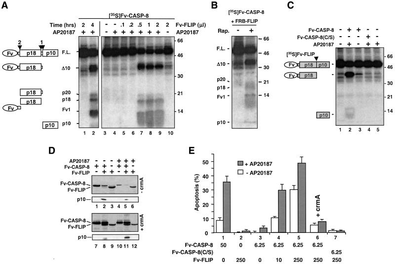
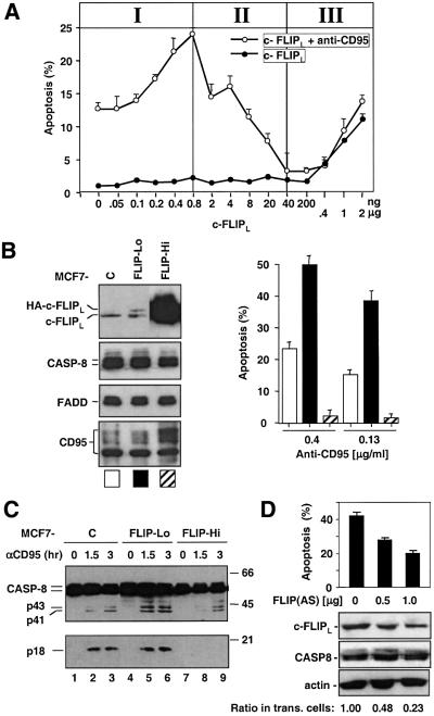
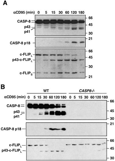
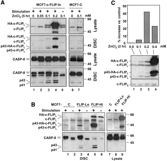
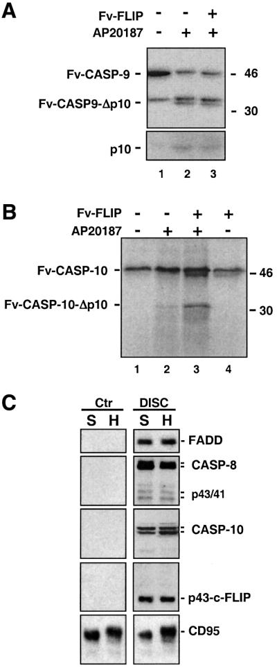
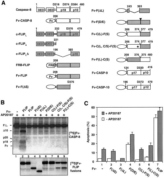
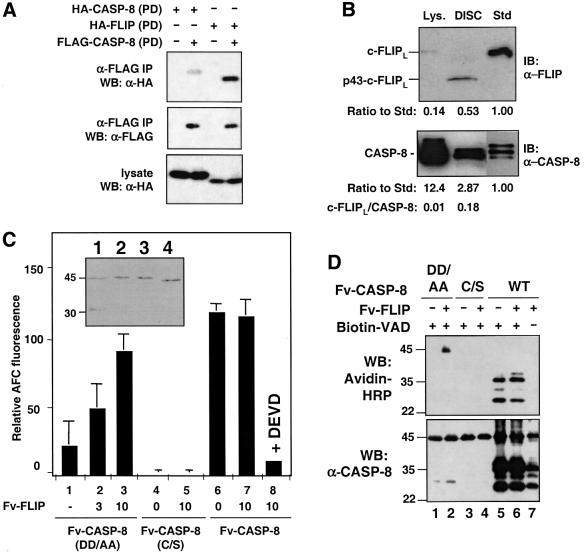
Activation of the caspase cascade is a pivotal step in apoptosis and can occur via death adaptor-mediated homo-oligomerization of initiator procaspases. Here we show that c-FLIP(L), a protease-deficient caspase homolog widely regarded as an apoptosis inhibitor, is enriched in the CD95 death-inducing signaling complex (DISC) and potently promotes procaspase-8 activation through hetero-dimerization. c-FLIP(L) exerts its effect through its protease-like domain, which associates efficiently with the procaspase-8 protease domain and induces the enzymatic activity of the zymogen. Ectopic expression of c-FLIP(L) at physiologically relevant levels enhances procaspase-8 processing in the CD95 DISC and promotes apoptosis, while a decrease of c-FLIP(L) expression results in inhibition of apoptosis. c-FLIP(L) acts as an apoptosis inhibitor only at high ectopic expression levels. Thus, c-FLIP(L) defines a novel type of caspase regulator, distinct from the death adaptors, that can either promote or inhibit apoptosis.


Figures

None
Fig. 1. c-FLIPL enhances caspase-8 activation upon induced proximity. (A) The c-FLIPL protease-like domain enhances caspase-8 processing. A 1 µl aliquot of in vitro translated, 35S-labeled Fv-CASP-8 was treated with 100 nM AP20187 for the indicated time (lanes 1 and 2), or treated with vehicle (–) or 100 nM AP20187 (+) for 2 h in the presence of the indicated amounts of in vitro translated, non-radioisotope-labeled Fv-FLIP (lanes 3–10). The reaction mixtures were then resolved by SDS–PAGE, and 35S-labeled products were visualized by autoradiography. The deduced domain structures of the marked bands are shown on the left and the order of processing is marked above. Molecular weight standards (in kDa) are shown on the right. (B) Enhancement of caspase-8 processing by c-FLIPL is due to their hetero-dimerization. A 1 µl aliquot of [35S]Fv-CASP-8 was treated with 1 mM rapamycin (Rap.) for 8 h in the presence of 1 µl of unlabeled FRB-FLIP. The products were analyzed as in (A). No processing was observed in the absence of FRB-FLIP (not shown). Domain structures are shown on the left and molecular weight standards on the right. (C) Dimerization induces processing of Fv-FLIP by caspase-8. A mixture of 1 µl of [35S]Fv-FLIP and 1 µl of unlabeled Fv-CASP-8 or Fv-CASP-8(C360S) was treated with or without AP20187 for 2 h and analyzed as in (A). The deduced domain structures are shown on the left. (D) Caspase-8 processing in transfected cells. 293 cells were transfected with the indicated combinations of Fv-CASP-8 (1 µg), Fv-FLIP (1 µg) and pRK5-CrmA (2 µg). At 13 h after transfection, cells were treated with vehicle (–) or 50 nM AP20187 (+) for 5 h. Cell extracts were analyzed by immunoblotting analysis using an anti-FLAG antibody. (E) Fv-FLIP enhances the cell death activity of caspase-8 in mammalian cells. HeLa cells were transfected with the indicated combinations of Fv constructs (in ng) and pRK5-crmA (1.5 µg, lane 6), together with pCMV-lacZ. At 6 h after transfection, cells were incubated with AP20187 (final concentration 125 nM), FK506 (200 nM) and z-DEVD (5 µM) for 10 h and scored for apoptosis.
None
Fig. 2. c-FLIPL functions as an activator for CD95-mediated apoptosis. (A) Dosage-dependent effect of c-FLIPL on CD95-mediated apoptosis. HeLa cells seeded in 6-well plates were transfected with the indicated amounts of c-FLIPL/pcDNA3 construct plus the vector DNA to make the total amount of DNA constant. After 16 h, cells were treated with CH11 (75 ng/ml) plus cycloheximide (1 µg/ml) for 6 h and scored for apoptosis. (B) CD95-mediated apoptosis in MCF7-CD95 cells stably expressing c-FLIPL. Left: expression of c-FLIPL, caspase-8, FADD and CD95 in the MCF7-CD95 cells expressing no exogenous c-FLIPL (MCF7-C) or stably expressing low (MCF7-FLIP-Lo) or high levels (MCF7-FLIP-Hi) of c-FLIPL. The exogenous c-FLIPL (HA-c-FLIPL) migrated more slowly than the endogenous c-FLIPL on SDS–PAGE due to the tags. Right: MCF7-CD95 cells were treated with the indicated concentrations of anti-APO-1 plus protein A (5 ng/ml) for 3 h and scored for apoptosis. These cells were similarly sensitive to staurosporine-induced apoptosis (data not shown). (C) Activation of overall caspase-8 in the MCF7-CD95 cells expressing different levels of c-FLIPL. Different MCF7 cells were treated with anti-APO-1 as in (B) for the indicated times, and whole-cell lysates were analyzed by immunoblotting with the anti-caspase-8 mAb C15, which recognized a region in the large subunit. No p18 band was detected in MCF7-FLIP-Hi cells even after a much longer exposure. (D) Endogenous c-FLIPL is required for CD95-mediated apoptosis. HeLa cells cultured in 12-well plates were transfected with the indicated amount of the c-FLIPL antisense plasmid plus 0.15 µg of pEGFP-N3. The total amount of DNA used for transfection was made constant using vector DNA. Top panel: 24 h after transfection, cells were treated with anti-APO-1 (0.4 ng/ml) plus protein A (5 ng/ml) for 4 h, and green fluorescent protein (GFP)-positive cells were scored for apoptosis. Without CD95 stimulation, the background cell death in each condition was approximately the same and <5%. Bottom panels: expression levels of endogenous c-FLIPL, caspase-8 and actin detected at 24 h post-transfection. The ratio of c-FLIP proteins in different lanes was determined using a Bio-Rad Densitometer GS-700, which was then adjusted according to the transfection efficiency (∼70% as judged by EGFP expression) to obtain the ratio of c-FLIPL in transfected (trans.) cells.
None
Fig. 3. Processing of procaspase-8 and c-FLIPL during CD95-mediated apoptosis. (A) Processing of caspase-8 and c-FLIPL in MCF7-CD95 cells. MCF7-CD95 cells were treated with anti-APO-1 (0.5 µg/ml) plus protein A (10 ng/ml) for the indicated times. Whole-cell lysates were analyzed by immunoblotting with anti-caspase-8 (top and middle) or anti-c-FLIP (bottom) antibody. Molecular weight standards are shown on the right. (B) Processing of c-FLIPL requires caspase-8 activity. Wild-type or caspase-8 deficient Jurkat cells were treated with anti-APO-1 and the cell lysates were analyzed as in (A). The small amount of c-FLIPL processing after prolonged treatment was probably caused by caspase-10, which is also recruited to the DISC (Figure 5C).
None
Fig. 4. c-FLIPL enhances caspase-8 activation in the DISC. (A) Effects of c-FLIPL on caspase-8 processing in the DISC in MCF7-CD95 cells inducibly expressing c-FLIPL. MCF7-FLIP-In cells were treated with the indicated concentration of ZnCl2 for 5 h. Cells were then stimulated with anti-APO-1 (+) or left untreated (–), and DISC complexes were isolated. Cell lysates and DISC were subjected to immunoblotting analysis using anti-c-FLIP (top two panels) or anti-caspase-8 antibody (bottom two panels). **IgG heavy chain. (B) Caspase-8 processing in MCF7 transfectants stably expressing c-FLIPL. Different MCF7 cells were stimulated with anti-APO-1 or left untreated. Cell lysates and DISC were analyzed by immunoblotting with anti-c-FLIP (top panels) or anti-caspase-8 (bottom panels) antibody. (C) c-FLIPL increases caspase-8 activity in the DISC. A total of 2 × 107 MCF7-c-FLIP-In cells were treated with ZnCl2 for 2 h, washed and kept without ZnCl2 for another 4 h at 37°C. The DISC was isolated and the caspase-8 activity in the DISC was assayed using IETD-AFC. The data shown are representative of three independent experiments.
None
Fig. 5. Specific effect of c-FLIPL on caspase-8 and -10 and identification of caspase-10 as a DISC component. (A and B) Fv-FLIP enhances caspase-10 but not caspase-9 processing. A 1 µl aliquot of [35S]Fv-CASP-9 (A) or [35S]Fv-CASP-10 (B) was treated with 100 nM AP20187, in the presence or absence of 1 µl of unlabeled Fv-FLIP. The reaction mix was analyzed as in Figure 1A. Domain structures are shown on the left, and molecular weight standards on the right. (C) Identification of caspase-10 as a DISC component. CD95 was immunoprecipitated from stimulated (DISC) or unstimulated (Ctr) SKW6.4 (S) and H9 (H) cells. CD95 and the associated proteins were analyzed by immunoblotting using appropriate monoclonal antibodies.
None
Fig. 6. The entire protease-like domain is required for c-FLIPL function. (A) Schematic structures of caspase-8, c-FLIPL, c-FLIPS and various constructs used in this study. DED, death effector domain. Amino acids present in each construct are labeled. Regions of c-FLIPL and caspase-8 in the chimeric constructs are indicated by shaded and open boxes, respectively. The domains that form the mature caspases as well as the corresponding domain on c-FLIPL (p10, p18, etc.) are marked. Fv, a derivative of FK506-binding protein (Clackson et al., 1998); FRB, the minimal rapamycin- binding domain of the FKBP–rapamycin-associated protein (FRAP) (Chen et al., 1995); asterisk, caspase-8 active site Cys360 to serine mutation; arrowhead, c-FLIPL processing site Asp376 to alanine mutation. (B) Capase-8 processing induced by c-FLIPL mutants and c-FLIPL:caspase-8 chimeras. Top panel: 1 µl of [35S]Fv-CASP-8 was treated with or without AP20187, in the presence of 1 µl of the indicated unlabeled Fv fusion proteins. Deduced domain structures are shown on the left. Bottom panel: the Fv fusion proteins used in the top panel were labeled with [35S]methionine to confirm equal expression of protein. (C) Effects of c-FLIPL mutants and c-FLIPL:caspase-8 chimeras on caspase-8-induced apoptosis. HeLa cells were transfected with Fv-CASP-8 (25 ng) and the indicated Fv-FLIP mutants (500 ng), and apoptosis was determined as in Figure 1E.
None
Fig. 7. The c-FLIPL protease-like domain induces the enzymatic activity of the caspase-8 zymogen. (A) The c-FLIPL protease-like domain interacts with the caspase-8 protease domain more efficiently than the caspase-8 protease domain does with itself. 293 cells were transfected with HA-tagged caspase-8 protease domain (PD) or HA-tagged c-FLIPL protease-like domain together with FLAG-tagged caspase-8-PD. The cell lysates were immunoprecipitated with an anti-FLAG antibody. The immunoprecipitated proteins and the lysates were analyzed by immunoblotting as indicated. (B) Ratios of c-FLIPL versus caspase-8 in cell lysate and in the DISC. The DISC complexes and whole-cell lysates (lys.) were made from 3 × 106 MCF7-CD95 cells. Thirty percent of these DISC complexes and 0.3% of the lysate (lys.) were resolved by SDS–PAGE together with a c-FLIPL (top) or a caspase-8 (bottom) standard (Std), and analyzed by immunoblotting (IB) with the indicated antibodies. For making protein standards, HA-tagged c-FLIPL and caspase-8 proteins were generated by in vitro translation. The relative concentration of these two proteins was determined by anti-HA immunoblotting, and equal amounts of these two proteins were used. The ratio of c-FLIPL/caspase-8 in the DISC was higher when the expression level of c-FLIPL increased. In the MCF7-CD95 cells stably expressing low and high levels of c-FLIPL, the ratio is ∼1:2 and 10:1, respectively (Figure 4B, lanes 3 and 5). (C and D) Fv-FLIP enhances the caspase activity of non-cleavable Fv-CASP-8(DD/AA). (C) Recombinant Fv-CASP-8 fusion proteins (100 ng) were incubated with the indicated amount of Fv-FLIP protein (in ng). Caspase-8 activity was measured using IETD-AFC. The data shown were representative of three independent experiments done in duplicate or triplicate. The insert shows analysis of recombinant Fv-CASP-8 (1), Fv-CASP-8(DD/AA) (2), Fv-CASP(C/S) (3) and Fv-FLIP (4) purified from bacteria. About 100 ng of each protein was resolved by SDS–PAGE and visualized by Coomassie Blue staining. (D) Fv-CASP-8 proteins were labeled with biotin-DEVD in the presence or absence of Fv-FLIP. The mixes were then resolved by SDS–PAGE and immunoblotted with either avidin–HRP (top) or anti-caspase-8 mAb C15 (bottom).

References

    1. Chang H.Y. and Yang,X. (2000) Proteases for cell suicide: functions and regulation of caspases. Microbiol. Mol. Biol. Rev., 64, 821–846. - PMC - PubMed
    1. Chen J., Zheng,X.F., Brown,E.J. and Schreiber,S.L. (1995) Identification of an 11-kDa FKBP12–rapamycin-binding domain within the 289-kDa FKBP12–rapamycin-associated protein and characterization of a critical serine residue. Proc. Natl Acad. Sci. USA, 92, 4947–4951. - PMC - PubMed
    1. Clackson T. et al. (1998) Redesigning an FKBP–ligand interface to generate chemical dimerizers with novel specificity. Proc. Natl Acad. Sci. USA, 95, 10437–10442. - PMC - PubMed
    1. Goltsev Y.V., Kovalenko,A.V., Arnold,E., Varfolomeev,E.E., Brodianskii,V.M. and Wallach,D. (1997) CASH, a novel caspase homologue with death effector domains. J. Biol. Chem., 272, 19641–19644. - PubMed
    1. Griffith T.S., Chin,W.A., Jackson,G.C., Lynch,D.H. and Kubin,M.Z. (1998) Intracellular regulation of TRAIL-induced apoptosis in human melanoma cells. J. Immunol., 161, 2833–2840. - PubMed

Publication types