. 2002 Jul 15;21(14):3782-93.
doi: 10.1093/emboj/cdf361.

Growth factors can activate ATF2 via a two-step mechanism: phosphorylation of Thr71 through the Ras-MEK-ERK pathway and of Thr69 through RalGDS-Src-p38

Affiliations

Growth factors can activate ATF2 via a two-step mechanism: phosphorylation of Thr71 through the Ras-MEK-ERK pathway and of Thr69 through RalGDS-Src-p38

D Margriet Ouwens et al. EMBO J. .

Abstract

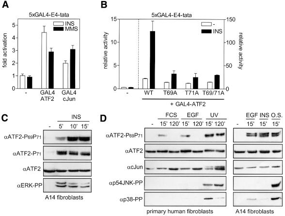
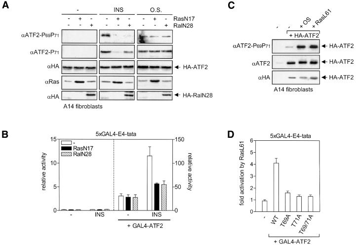
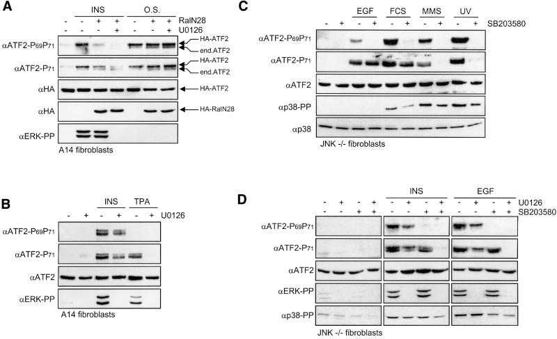
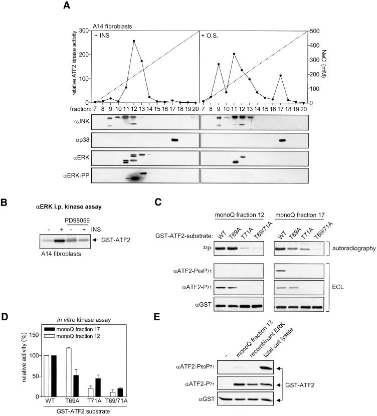
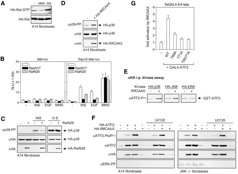
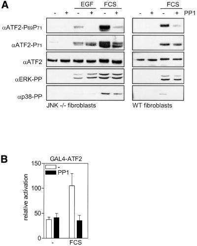
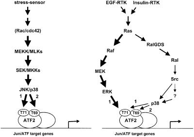
Transcription factor ATF2 regulates gene expression in response to environmental changes. Upon exposure to cellular stresses, the mitogen-activated proteinkinase (MAPK) cascades including SAPK/JNK and p38 can enhance ATF2's transactivating function through phosphorylation of Thr69 and Thr71. How ever, the mechanism of ATF2 activation by growth factors that are poor activators of JNK and p38 is still elusive. Here, we show that in fibroblasts, insulin, epidermal growth factor (EGF) and serum activate ATF2 via a so far unknown two-step mechanism involving two distinct Ras effector pathways: the Raf-MEK-ERK pathway induces phosphorylation of ATF2 Thr71, whereas subsequent ATF2 Thr69 phosphorylation requires the Ral-RalGDS-Src-p38 pathway. Cooperation between ERK and p38 was found to be essential for ATF2 activation by these mitogens; the activity of p38 and JNK/SAPK in growth factor-stimulated fibroblasts is insufficient to phosphorylate ATF2 Thr71 or Thr69 + 71 significantly by themselves, while ERK cannot dual phosphorylate ATF2 Thr69 + 71 efficiently. These results reveal a so far unknown mechanism by which distinct MAPK pathways and Ras effector pathways cooperate to activate a transcription factor.


Figures

None
Fig. 1. Insulin activates ATF2 through phosphorylation of Thr69 and Thr71. (A) Insulin efficiently enhances the transactivating capacity of ATF2. A14 cells were transiently transfected with 2 µg of 5×GAL4-E4-luciferase reporter in the presence or absence of 2 µg of pRSV-GAL4-cJun-N, pRSV-GAL4-ATF2-N or an empty expression vector. At 20 h after transfection, the cells were incubated for 16 h with 10 nM insulin or 1 mM MMS. Transactivation by GAL4-cJun and GAL4-ATF2 in the absence of insulin and MMS was 2.4- and 46-fold, respectively. For comparison, only the fold activation (mean ± SD) by insulin and MMS is depicted, which represents the ratio between relative luciferase activity in the presence and absence of insulin or MMS. (B) Insulin-induced transactivation by ATF2 requires Thr69 and Thr71. A14 cells were transiently transfected with 0.5 µg of 5×GAL4-E4-luciferase reporter plasmid in the presence or absence of 2 µg of the indicated pC2-GAL4-ATF2 expression vectors, encoding either the wild-type (wt) ATF2 transactivation domain, or the corresponding domain in which Thr69 (T69A), Thr71 (T71A) or both (T69/71A) are replaced by alanine. At 6 h after transfection, the cells were stimulated for 16 h with 10 nM insulin. The relative activity is the enhancement of promoter activity by the various GAL4-ATF2 fusion proteins in the absence and presence of insulin, and is presented as the mean ± SD of two independent experiments performed in triplicate. Note the different scaling of the left and right y-axis. (C) Insulin-induced ATF2 Thr71 and Thr69 + 71 phosphorylation. Serum-starved A14 cells were stimulated with 10 nM insulin for the indicated times. Total cell extracts (30 µg of protein) were analyzed by SDS–PAGE and immunoblotting. Ponceau S staining confirmed that the filters contained equal amounts of protein extracts. The faster migrating bands seen by the phospho-specific ATF2 antibodies seem to represent shorter, alternatively spliced, ATF2 products (Georgopoulus et al., 1992). (D) Mitogen-induced ATF2 Thr69 + 71 phosphorylation does not correlate with JNK Thr183/Tyr185 and p38 Thr180/Tyr182 phosphorylation in A14 and primary human VH10 fibroblasts. Serum-starved cells were stimulated for the indicated times with 10 nM EGF, 10 nM insulin, 500 mM NaCl (O.S.), 10% FCS or 30 J/m2 UVC. Total cell extracts (30 µg of protein) were analyzed for the levels of the indicated proteins by SDS–PAGE and subsequent immunoblotting.
None
Fig. 2. Ras-dependent growth factor-induced activation of ATF2. (A) Inhibition of insulin-induced ATF2 phosphorylation by dominant-negative Ras and Ral. A14 cells were transiently transfected with 0.5 µg of pMT2-HA-ATF2 in the presence or absence of 2 µg of pRSV-RasN17, pMT2-HA-RalN28 or an empty expression vector. Fugene reagent was used in order to obtain high levels of transfection efficiency (>40%). At 24 h after transfection, the cells were stimulated with either 10 nM insulin (15 min) or 500 mM NaCl (O.S.) (15 min). A 30 µg aliquot of total cell extract was analyzed by SDS–PAGE/immunoblotting. Ectopically expressed RasN17, HA-ATF2 and HA-RalN28 were detected using monoclonal Y13-259 Ras and HA antibodies, respectively. Comparison of the extracts of mock-transfected cells with those of HA-ATF2 transfected cells verified that only exogenous HA-ATF-2 is detected on the exposures shown [data not shown; compare with (C) and Figure 5F]. (B) Insulin-induced GAL4-ATF2-dependent transactivation is inhibited by RasN17 and RalN28. A14 cells were transiently transfected with 0.5 µg of 5×GAL4-E4-luciferase reporter together with 2 µg of expression vectors for RasN17 and RalN28, or an empty control vector, in the presence or absence of either 1 µg of pRSV-GAL4-ATF2-N or an empty expression vector. Cells were serum starved for 24 h, followed by stimulation with 10 nM insulin for 14 h. Depicted is the enhancement of promoter activity by GAL4-ATF2 in the absence and presence of insulin and/or the inhibitors (mean ± SD). Note the different scaling of the left and right y-axis. (C) Active RasL61 induces ATF2 Thr69 + 71 phosphorylation. A14 cells were kept untreated (–), or transfected with 0.5 µg of pMT2-HA-ATF2 in the presence or absence of 2 µg of RasL61 expression vector, or an empty vector (–). Fugene reagent was used in order to obtain high levels of transfection efficiency (>40%). At 24 h after transfection, cells were stimulated with NaCl (O.S.), as described for Figure 1D, when indicated, and total cell lysates were prepared and analyzed by SDS–PAGE and immunoblotting. (D) Active RasL61 enhances transactivation by ATF2 via ATF2 Thr69 and Thr71. A14 cells were transiently transfected with 2 µg of 5×GAL4-E4-luciferase reporter plasmid together with 2 µg of pRSV-GAL4-ATF2 expression vectors containing full-length (wt) ATF2, or the corresponding domain in which Thr69 (T69A), Thr71 (T71A) or both (T69/71A) are replaced by alanine. In addition to these GAL4 fusion constructs, 3 µg of pRSV-RasL61, or an empty expression vector, was co-transfected. At 40 h after transfection, cells were harvested and analyzed for luciferase activity. The fold activation depicted represents the ratio between luciferase activity (mean ± SD) in the presence and absence of RasL61.
None
Fig. 3. Differential phosphorylation of ATF2 Thr71 and Thr69 by the Raf–MEK and RalGDS–Ral effector pathways of Ras. (A) Inhibition of insulin-induced ATF2 phosphorylation by RalN28 and the MEK inhibitor U0126. A14 cells were transfected and stimulated with either insulin or NaCl (O.S.) as described for Figure 2A. Where indicated, the cells were pre-treated for 30 min with 10 µM U0126. A 30 µg aliquot of total cell extract was analyzed by SDS–PAGE/immunoblotting. The positions of the exogenously expressed HA-ATF2 and the faster migrating endogenous (end.) ATF2 are indicated on the right. (B) Activation of the Raf–MEK pathway induces ATF2 Thr71 mono-phosphorylation. Serum-starved A14 cells were incubated for 30 min in the absence or presence of 10 µM U0126 prior to treatment with 10 nM insulin or 100 nM TPA. Total cell extracts (30 µg of protein) were prepared after 15 min, and analyzed by SDS–PAGE/immunoblotting. (C) Differential effects of SB203580 on ATF2 Thr71 phosphorylation induced by growth factors and stresses. Serum-starved JNK1+2–/– fibroblasts were incubated for 30 min in the absence or presence of 5 µM SB203580 prior to treatment with 10 nM EGF, 20% FCS, 30 J/m2 UVC or 1 mM MMS as indicated. Total cell extracts (30 µg of protein) were prepared after 15 min (except for MMS: 2 h), and analyzed by SDS–PAGE and subsequent immunoblotting. (D) Differential effects of SB203580 and U0126 on ATF2 Thr71 and Thr69 + 71 phosphorylation. Serum-starved JNK1+2–/– fibroblasts were treated for 30 min with 5 µM SB203580 and/or 10 µM U0126 prior to stimulation for 15 min with 10 nM insulin or 10 nM EGF as indicated. Total cell extracts (30 µg of protein) were analyzed by SDS–PAGE and immunoblotting.
None
Fig. 4. Insulin- and EGF-induced ATF2 Thr71 mono-phosphorylation is mediated by ERK. (A) The main insulin-induced ATF2 N-terminal kinase activity co-purifies with ERK1/2 after MonoQ anion-exchange chromatography. Total cell lysates from A14 cells treated for 15 min with either 10 nM insulin or 500 mM NaCl (O.S.) were separated on a MonoQ column using a linear gradient of NaCl (dotted line). Fractions were analyzed for in vitro ATF2 kinase activity (filled circles) as described in Materials and methods, and for the presence of JNK, ERK1/2 and p38 by SDS–PAGE and immunoblotting. (B) Insulin induces ERK-associated ATF2 N-terminal kinase activity. Serum-starved A14 cells were either untreated or treated for 15 min with 10 nM insulin, with or without 15 min pre-treatment with 20 µM PD98059, as indicated. Total cell lysates were immunoprecipitated with antibodies that recognize both ERK1 and ERK2, and subsequently assayed for ATF2-kinase activity. (C) Insulin-induced ERK and p38 activities differ in their ATF2 Thr69 and Thr71 kinase activities. Partially purified ERK and p38 preparations from insulin-stimulated A14 cell extracts (MonoQ fractions 12 and 17, respectively) were analyzed for in vitro kinase activity, using either wild-type (wt) or mutant GST–ATF2 fusion proteins in which Thr69 (T69A), Thr71 (T71A) or Thr69 + 71 (T69/71A) were replaced by alanine. The phosphorylation state subsequently was monitored by SDS–PAGE followed by autoradiography and immunoblotting using phospho-specific antibodies followed by enhanced chemiluminescence (ECL). (D) Quantification of 32P incorporation into GST–ATF2 by MonoQ fractions 12 and 17 in in vitro kinase assays as described in (C). The relative activity (mean ± SD) shown represents the 32P incorporation in the various mutant GST–ATF2 substrates relative to that in the wild-type GST–ATF2 protein (set at 100% for both fractions 12 and 17). (E) Recombinant active ERK only phosphorylates ATF2 Thr71 efficiently. Recombinant ERK (10 U; Calbiochem) was compared with MonoQ fraction 13 and total cell lysate from insulin-treated A14 cells for its ATF2 kinase potential using GST–ATF2 as a substrate. The phosphorylation state of GST–ATF2 Thr69 + 71 and GST–ATF2 Thr71 subsequently was monitored by SDS–PAGE/immunoblotting using phospho-specific antibodies. Equal loading of the GST–ATF2 substrate was verified by reprobing the filters with GST antibodies.
None
Fig. 5. The RalGDS–Ral pathway mediates insulin- and EGF-induced activation of ATF2-dependent gene expression. (A) The effects of insulin and MMS on Ral activity. A14 cells were transiently transfected with 3 µg of pMT2-HA-Ral. At 24 h after transfection, the cells were serum-starved overnight followed by stimulation with either 10 nM insulin (15 min) or 1 mM MMS (2 h). Total cell extracts (750 µg of protein) were incubated with 15 µg of GST–RalBD pre-coupled to glutathione beads to recover GTP-bound Ral. Beads were washed extensively, and collected Ral was detected by immunoblotting with HA antibody. (B) Insulin- and EGF-induced activation of ATF2-dependent transcription is inhibited by RasN17 and RalN28. A14 cells were transiently transfected with 2 µg of either the cJun–ATF2-dependent luciferase reporter 5×jun2-tata or the tata-luciferase control, in the presence or absence of 2 µg of expression vectors for RasN17 and RalN28, or an empty control vector. At 20 h after transfection, the cells were stimulated for 6 h with 10 nM insulin or 1 mM MMS. Depicted is the relative luciferase activity (RLU) ± SD. (C) Dominant-negative Ral inhibits insulin-induced p38 phosphorylation. A14 cells were transiently transfected with 0.5 µg of pMT2-HA-p38 in the presence or absence of 1.5 µg of pMT2-HA-RalN28, or an empty expression vector as described in Figure 2A. Subsequently, the cells were serum-starved and treated with either 10 nM insulin or 500 mM NaCl (O.S.). Total cell extracts were prepared after 15 min, and analyzed by SDS–PAGE/immunoblotting. For better comparison, a relatively short exposure of osmotic shock-induced HA-phospho-p38 is shown. (D) Activation of Ral by RlfCAAX induces p38 phosphorylation. A14 cells were transfected with 0.5 µg pMT2-HA-p38 in the presence or absence of 0.125 µg of HA-RlfCAAX, or an empty vector (–) as described above. At 24 h after transfection, the cells were serum-starved and, after an additional 24 h, total cell lysates were prepared and analyzed by SDS–PAGE and immunoblotting. (E) Activation of Ral by RlfCAAX induces p38 and JNK kinase activity. A14 cells were transfected with 0.5 µg of expression vectors encoding HA-tagged p38, JNK or ERK, respectively, in the presence or absence of 0.125 µg of HA-RlfCAAX expression vector, or an empty vector (–) as described above. At 24 h after transfection, the cells were serum-starved and, after an additional 24 h, total cell lysates were prepared. Lysates were immunoprecipitated with an HA antibody, after which HA-associated ATF2 Thr71 kinase activity was measured using GST–ATF2 as substrate (see Materials and methods). (F) Activation of Ral by RlfCAAX induces ATF2 Thr69 + 71 phosphorylation. A14 and JNK–/– cells were left untreated (–) or transfected with 0.5 µg of pMT2-HA-ATF2 in the presence or absence of 0.125 µg of RlfCAAX expression vector. Fugene reagent was used in order to obtain high levels of transfection efficiency (>40%). At 24 h after transfection, cells were serum-starved overnight, and incubated for a further 24 h in the presence or absence of 10 µM U0126 prior to preparation of cell lysates and analysis by SDS–PAGE and immunoblotting. Note that HA-ATF2 and HA-RlfCAAX (detected by the HA antibody) have nearly the same molecular weight. (G) RlfCAAX enhances transactivation by ATF2 via ATF2 Thr69 and Thr71. A14 cells were transiently transfected with 2 µg of 5×GAL4-E4-luciferase reporter plasmid together with 2 µg of pRSV-GAL4-ATF2 expression vectors containing full-length (wt) ATF2, or the corresponding domain in which Thr69 (T69A), Thr71 (T71A) or both (T69/71A) are replaced by alanine. In addition to these GAL4 fusion constructs, 3 µg of pMT2-RlfCAAX, or an empty expression vector was co-transfected. At 40 h after transfection, cells were harvested and analyzed for luciferase activity. The fold activation depicted represents the ratio between luciferase activity in the presence and absence of RlfCAAX. Values represent the mean ± SD.
None
Fig. 6. Src-like proteins are required for growth factor-induced ATF2 Thr69 phosphorylation. (A) Serum-starved JNK1+2–/– and wild-type 3T3 fibroblasts were incubated for 30 min in the absence or presence of 10 µM PP1 prior to stimulation with 10 nM EGF, or 20% FCS as indicated. Total cell extracts (30 µg of protein) were prepared after 15 min, and analyzed by SDS–PAGE/immunoblotting. (B) Serum- induced transactivation by GAL4-ATF2 is inhibited by PP1. 3T3 fibroblasts were transiently transfected with 0.5 µg of 5×GAL4-E4- luciferase reporter plasmid in the presence or absence of 2 µg of pC2-GAL4-ATF2 expression vector. At 24 h after transfection, the cells were serum-starved overnight prior to treatment for 14 h with 20% FCS in the presence or absence of 10 µM PP1. The relative activation is the enhancement of promoter activity by the GAL4-ATF2 fusion protein in the absence and presence of serum, and is presented as the mean ± SD of two independent experiments performed in triplicate.
None
Fig. 7. Proposed model for the order of events leading to ATF2 Thr69 + 71 phosphorylation by growth factors and stresses. For details, see Discussion.

Similar articles

Cited by

References

    1. Angel P., Hattori,K., Smeal,T. and Karin,M. (1988) The jun proto-oncogene is positively autoregulated by its product, Jun/AP-1. Cell, 55, 875–885. - PubMed
    1. Bakiri L., Lallemand,D., Bossy-Wetzel,E. and Yaniv,M. (2000) Cell cycle-dependent variations in c-Jun and JunB phosphorylation: a role in the control of cyclin D1 expression. EMBO J., 19, 2056–2068. - PMC - PubMed
    1. Bar-Sagi D. and Hall,A. (2000) Ras and Rho GTPases: a family reunion. Cell, 103, 227–238. - PubMed
    1. Beier F., Lee,R.J., Taylor,A.C., Pestell,R.G. and LuValle,P. (1999) Identification of the cyclin D1 gene as a target of activating transcription factor 2 in chondrocytes. Proc. Natl Acad. Sci. USA, 96, 1433–1438. - PMC - PubMed
    1. Beier F., Taylor,A.C. and LuValle,P. (2000) Activating transcription factor 2 is necessary for maximal activity and serum induction of the cyclin A promoter in chondrocytes. J. Biol. Chem., 275, 12948–12953. - PubMed

Publication types

MeSH terms