. 2002 Jul 15;21(14):3816-28.
doi: 10.1093/emboj/cdf376.

Site-specific cross-linking analyses reveal an asymmetric protein distribution for a box C/D snoRNP

Affiliations

Site-specific cross-linking analyses reveal an asymmetric protein distribution for a box C/D snoRNP

Niamh M Cahill et al. EMBO J. .

Abstract

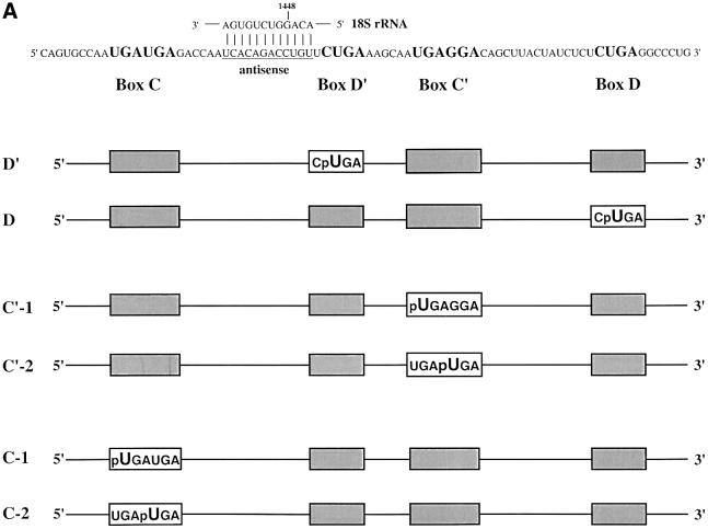
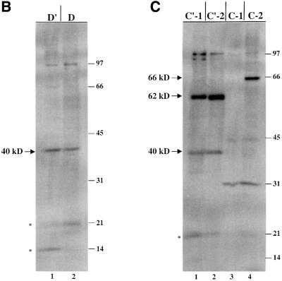
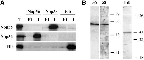
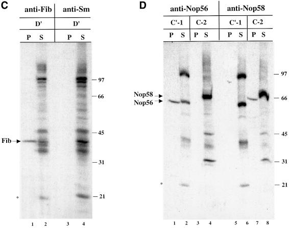
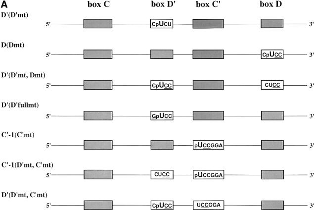
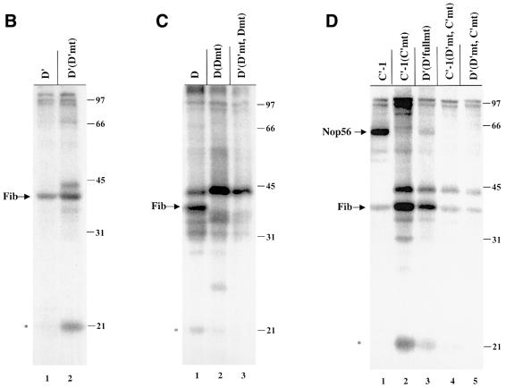
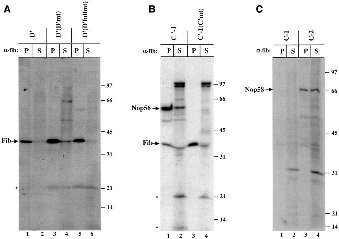
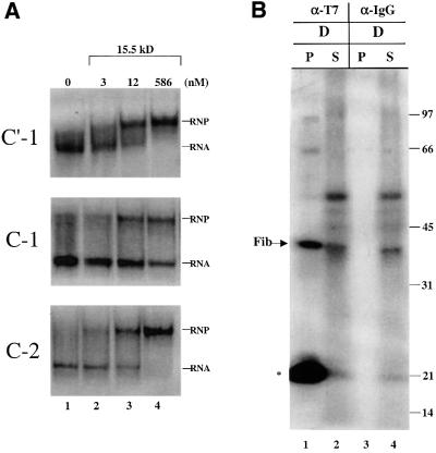
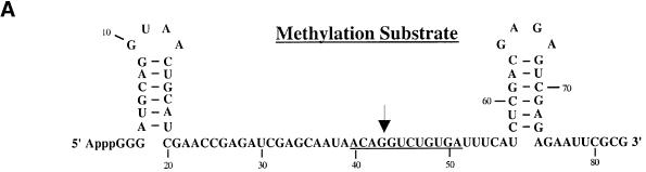
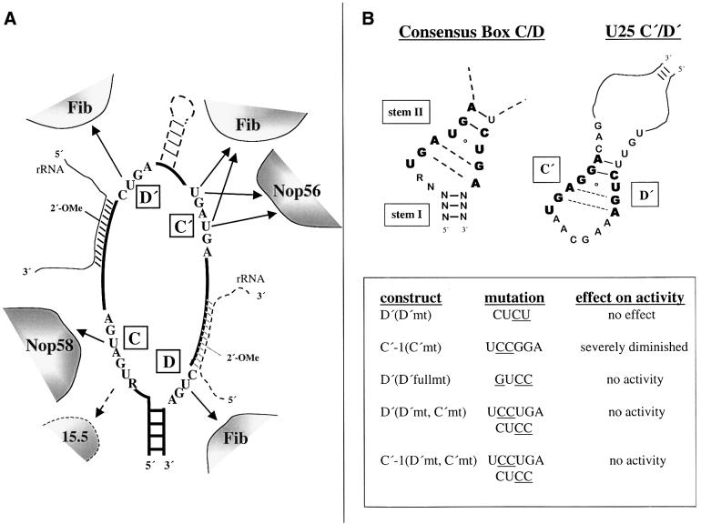
Methylation of the ribose 2'-hydroxyl, the most widespread modification of ribosomal and splicesomal RNAs, is guided by the box C/D class of small nucleolar RNAs (snoRNAs). Box C/D small nucleolar ribonucleoproteins (snoRNPs) contain four core proteins: fibrillarin, Nop56, Nop58 and 15.5 kDa. We constructed U25 snoRNAs containing a single photoactivatable 4-thiouridine at each U position within the conserved box C/D and C'/D' motifs. Proteins assembled on the snoRNA after injection into Xenopus oocyte nuclei were identified by cross-linking, and reconstituted particles characterized by functional rescue and mutational analyses. Our data argue that box C/D snoRNPs are asymmetric, with the C' box contacting Nop56 and fibrillarin, the C box interacting with Nop58, and the D and D' boxes contacting fibrillarin. No cross-link to 15.5 kDa was detected; its binding is disrupted by 4-thiouridine substitution in position 1 of the C box. Repositioning the guide sequence of U25 upstream of box D instead of D' revealed that both C/D motifs have the potential to function as guide centers, but, surprisingly, there was no alteration in protein cross-linking.


Figures

None
Fig. 1. U25 snoRNA constructs and cross-linking analyses. (A) The Xenopus U25 snoRNA sequence is shown, highlighting in bold the conserved box elements. Three additional nucleotides were added to the 5′ end and two nucleotides to the 3′ end to extend the terminal stem (Tycowski et al., 1996). The antisense region (underlined) is shown base-paired to the Xenopus 18S rRNA sequence it targets. For site- specific cross-linking, a uridine within one of the conserved sequence elements was replaced with a 4SU (U) with a single radioactive label immediately 5′ of the 4SU, denoted by p. Constructs are named based on the site of 4SU incorporation. (B and C) SDS–polyacrylamide gel analyses of proteins cross-linked to the U25 constructs. After assembly of the injected RNAs in oocyte nuclei, photocross-linking and RNase digestion, proteins were resolved by 15% SDS–PAGE. The 40, 62 and 66 kDa cross-linked proteins indicated with an arrow are identified in Figure 2. RNA bands are indicated by an asterisk. The positions of molecular weight markers (kDa) are shown on the right.
None
Fig. 2. Identification of the 40, 62 and 66 kDa cross-linked proteins. (A) Antibodies generated against Xenopus Nop56, Nop58 and fibrillarin were used for immunoprecipitation of in vitro translated, 35S-labeled Nop56, Nop58 and fibrillarin proteins. Immunoprecipitations with pre-immune and immune sera are shown. Total lanes (T) show 20% of the 35S-labeled protein included in the assays. (B) Immunoblot analysis using oocyte crude nuclear extracts was performed with the Nop56, Nop58 or fibrillarin antisera as indicated. (C and D) After injection of the D′, C′-1 or C-2 construct, the irradiated and RNase-digested nuclear extracts were subjected to immunoprecipitation prior to SDS–PAGE using anti-Sm (Y12) or the antisera characterized in (A) and (B). The resulting pellets (P) and 1/10 of the supernatants (S) were resolved on 12.5% SDS–polyacrylamide gels. The bands identified as fibrillarin (Fib), Nop56 and Nop58 are indicated with arrows. RNA bands are indicated with an asterisk. The positions of protein molecular weight markers are shown on the right.
None
Fig. 3. Proteins cross-linked to mutant U25 constructs. (A) Mutant constructs are named with reference to the site of 4SU incorporation, as in Figure 1A, followed by (in parentheses) the box element that has been mutated. Mutated sequences are underlined. (BD) The labeling of gels resolving proteins cross-linked to the mutant constructs is as in Figure 2B and C.
None
Fig. 4. Anti-fibrillarin antibodies co-immunoprecipitate cross-linked fibrillarin, Nop56 and Nop58. After injection of nuclei with the D′, D′(D′mt), D′(D′fullmt), C′-1, C′-1(C′mt), C-1 or C-2 construct, cross-linked samples were immunoprecipitated with the anti-fibrillarin monoclonal antibody (72B9). The pellets (P) and supernatants (S) were then RNase digested and equal amounts were resolved on 12.5% SDS–polyacrylamide gels. Labeling is as in Figures 2 and 3.
None
Fig. 5. The 15.5 kDa protein assembles on U25 constructs in the oocyte. (A) Substitution of 4SU at the C-1 position adversely affects the binding of the 15.5 kDa protein. The C′-1, C-1 or C-2 RNA was incubated either alone (lane 1) or in the presence of recombinant 15.5 kDa protein at the concentrations indicated (see Materials and methods). Complexes were resolved on an 8% non-denaturing polyacrylamide gel, with the RNA alone (RNA) and the shifted RNA complex (RNP) indicated. (B) Approximately 20 nuclei isolated under paraffin oil were co-injected with the D construct (21 fmol/nucleus) and a T7 epitope-tagged recombinant 15.5 kDa protein (12.5 µM; gift of T.Hirose). The nuclei were incubated, irradiated and immunoprecipitated with anti-T7 tag antibody or mouse IgG. Samples were RNase digested, and equal fractions of the pellets (P) and supernatants (S) were resolved on a 12.5% SDS–polyacrylamide gel as in Figures 2–4.
None
Fig. 6. U25 constructs reconstitute methylation activity in vivo. (A) The substrate used to assay the methylation activity of substituted U25 snoRNPs is shown, with underlining indicating sequences complementary to the U25 snoRNA and the arrow indicating the nucleotide targeted for methylation. (B) The methylation activity of reconstituted U25 snoRNPs was assessed by co-injecting a 4SU-containing U25 snoRNA construct with the methylation substrate into nuclei 18 h after oocytes were injected with an antisense U25 DNA oligonucleotide. Nuclear RNA was isolated 4 h later and incubated with the chimeric oligonucleotide chim-U25 and RNase H as indicated. Lanes 1 and 2 show the uninjected substrate incubated in the absence or presence of RNase H. In lanes 3 and 4, the endogenous U25 snoRNA was not depleted. The amount of residual full-length substrate (full) and the cleaved substrate (cleaved) provides a qualitative assessment of the extent of site-specific methylation. In lanes 19–30, the full-length product was more degraded than in lanes 1–18. The activity of each construct was reproduced in three separate experiments.
None
Fig. 7. The C′ box cross-links to Nop56 and the C box to Nop58, regardless of the location of the guide sequence. (A) U25 constructs were designed with the 15 nucleotide sequence between the C box and D′ box switched for the 19 nucleotide sequence between the C′ box and the D box. The constructs are schematized and named as in Figure 1A, with SW indicating switched. (B) Cross-linking and analyses of labeled proteins were as in Figure 1. (C) Reconstitution of methylation activity with the SW C′-1 construct was assessed as in Figure 6B.
None
Fig. 8. (A) Summary of the RNA–protein contacts identified by 4SU site-specific cross-linking. The position of the 15.5 kDa protein is indicated with a dashed arrow, since this reported interaction (Watkins et al., 2000) was not observed by cross-linking. The three contacts to fibrillarin do not imply three copies of the fibrillarin molecule; they could reside on one, two or three copies of the protein (see text). Whereas the antisense sequence is 5′ of the D′ box in wild-type U25, the antisense region was switched to be 5′ of the D box in the ‘SW’ constructs, with the rRNA substrate and additional sequences between boxes C′ and D′ found in some snoRNAs indicated by dashed lines. (B) Comparison of the C′/D′ sequences of the U25 snoRNA with those of the consensus box C/D motif (Watkins et al., 2000).

References

    1. Anderson C.W., Baum,P.R. and Gesteland,R.F. (1973) Processing of adenovirus 2-induced proteins. J. Virol., 12, 241–252. - PMC - PubMed
    1. Balakin A.G., Smith,L. and Fournier,M.J. (1996) The RNA world of the nucleolus: two major families of small RNAs defined by different box elements with related functions. Cell, 86, 823–834. - PubMed
    1. Bortolin M.-L., Ganot,P. and Kiss,T. (1999) Elements essential for accumulation and function of small nucleolar RNAs directing site-specific pseudouridylation of ribosomal RNAs. EMBO J., 18, 457–469. - PMC - PubMed
    1. Caffarelli E., Losito,M., Giorgi,C., Fatica,A. and Bozzoni,I. (1998) In vivo identification of nuclear factors interacting with the conserved elements of box C/D small nucleolar RNAs. Mol. Cell. Biol., 18, 1023–1028. - PMC - PubMed
    1. Cavaillé J., Nicoloso,M. and Bachellerie,J.P. (1996) Targeted ribose methylation of RNA in vivo directed by tailored antisense RNA guides. Nature, 383, 732–735. - PubMed

Publication types