. 2002 Jul 15;21(14):3829-40.
doi: 10.1093/emboj/cdf373.

Class I tyrosyl-tRNA synthetase has a class II mode of cognate tRNA recognition

Affiliations

Class I tyrosyl-tRNA synthetase has a class II mode of cognate tRNA recognition

Anna Yaremchuk et al. EMBO J. .

Abstract

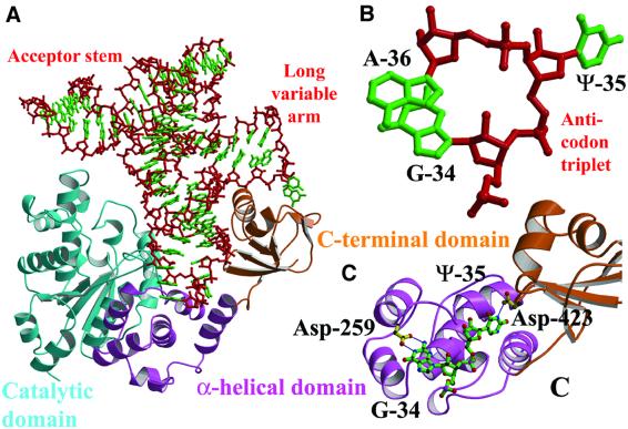
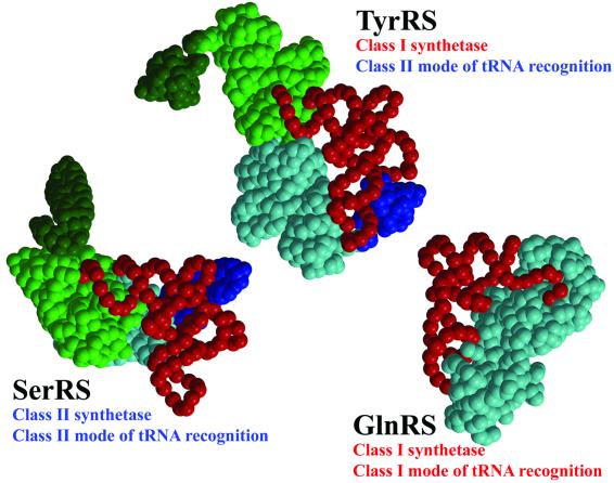
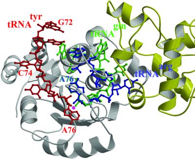
Bacterial tyrosyl-tRNA synthetases (TyrRS) possess a flexibly linked C-terminal domain of approximately 80 residues, which has hitherto been disordered in crystal structures of the enzyme. We have determined the structure of Thermus thermophilus TyrRS at 2.0 A resolution in a crystal form in which the C-terminal domain is ordered, and confirm that the fold is similar to part of the C-terminal domain of ribosomal protein S4. We have also determined the structure at 2.9 A resolution of the complex of T.thermophilus TyrRS with cognate tRNA(tyr)(G Psi A). In this structure, the C-terminal domain binds between the characteristic long variable arm of the tRNA and the anti-codon stem, thus recognizing the unique shape of the tRNA. The anticodon bases have a novel conformation with A-36 stacked on G-34, and both G-34 and Psi-35 are base-specifically recognized. The tRNA binds across the two subunits of the dimeric enzyme and, remarkably, the mode of recognition of the class I TyrRS for its cognate tRNA resembles that of a class II synthetase in being from the major groove side of the acceptor stem.


Figures

None
Fig. 1. Structure-based sequence alignment of tyrosyl-tRNA synthetase from T.thermophilus and B.stearothermophilus. The secondary structure of the TyrRSTT structure, calculated using DSSP (Kabsch and Sander, 1983), is superposed on top. Note the TyrRSTT-specific insertion between residues 302 and 330. The figure was prepared using ESPript (Gouet et al., 1999).
None
Fig. 2. Structure of T.thermophilus tyrosyl-tRNA synthetase and its tRNA complex. (A) Ribbon representation of the 2.0 Å resolution structure of T.thermophilus tyrosyl-tRNA synthetase complexed with tyrosinol. The catalytic domain is yellow (cyan), the α-helical domain red (pink) and the C-terminal domain green (orange), respectively, for each subunit in the dimer. The linkers between the α-helical and C-terminal domains, as well as some other loops involved in substrate binding, are disordered in this structure (see Table I). The small substrate tyrosinol is shown in solid-atom representation. (B) Same view, but shown as a stereoview, down the dimer 2-fold axis of the complex between T.thermophilus tyrosyl-tRNA synthetase, tRNAtyr(GΨA), tyrosinol and ATP. In this 2.9 Å resolution structure, all enzyme residues from 5–432 are visible, as are nucleotides 1–74 of the tRNA. Colours are as in (A), with the tRNA backbone in blue and the ATP and tyrosinol in solid-atom representation.
None
Fig. 3. Interactions between tyrosyl-tRNA synthetase and tRNAtyr. (A) The C-terminal domain (orange) binds in the elbow between the long variable arm and the anti-codon stem of the tRNA (red backbone, green bases). The anti-codon stem loop interacts with both the C-terminal domain and the α-helical domain (pink). The tRNA makes no contact with the catalytic domain of the same subunit (cyan). (B) The unusual conformation of the anti-codon triplet in which Ade-36 is stacked on Gua-34, while Psu-35 bulges out. (C) Base-specific interactions of Asp-259 from the α-helical domain with Gua-34 and Asp-423 from the C-terminal domain with Psu-35.
None
Fig. 4. Structure of tRNAtyr compared with that of tRNAser. (A) Comparison of the secondary structures of T.thermophilus tRNAtyr(GΨA) (left) and tRNAtyr(GGA) (right), highlighting differences, conserved in other prokaryotic organisms, that determine the orientation of the long variable arm. tRNAtyr nucleotides with only backbone contacts to TyrRSTT are shown in purple, those with only base contacts are shown in green and those with backbone and base contacts are shown in orange. (B) Comparison of the 3D structures of the base of the long variable arm in T.thermophilus tRNAtyr and T.thermophilus tRNAser (Biou et al., 1994), based on the structural alignment in (C). In tRNAser, Gua-20B is unpaired and stacks against the first base pair of the long variable arm, which comprises A45:U48-1 (top). In tRNAtyr, U48-1 is unpaired and stacks against the first base pair of the long variable arm, which comprises A20B:U48–2 (bottom). (C) View looking down the anticodon stem-loop of the structural alignment of tRNAtyr (blue) and tRNAser (red) based on superposition of 46 phosphates from the acceptor stem, D- and T-loops (r.m.s.d. = 1.16 Å). The tRNA cores have a very similar structure, but the variable arms project at an angle differing by ∼50°.
None
Fig. 5. Schematic diagrams showing different modes of tRNA recognition by TyrRS, SerRS and GlnRS. TyrRS (class I) and SerRS (class 2) both have a class II mode of tRNA recognition approaching the tRNA acceptor stem from the major groove side. GlnRS has a canonical class I mode of tRNA recognition approaching the tRNA acceptor stem from the minor groove side. Notably, both SerRS and TyrRS have long variable arm (class 2) tRNAs, are dimeric and have cross-subunit tRNA binding.
None
Fig. 6. Model of the 3′ CCA end of tRNAtyr in the active site of TyrRSTT. The C74-C75-A76 3′ extremity of tRNAtyr (red) is modelled in the active site of TyrRSTT by matching the predicted model of Labouze and Bedouelle (1989) to the experimental structure (this work) of tRNAtyr, which extends only to A73. Due to the close agreement of the position of the tRNAtyr in the experimental and predicted models, this is possible with only minor adjustments to avoid steric clashes with enzyme side-chains. The backbone conformation of the CCA and the positions of the bases are by no means necessarily correct. The aim is to show that the observed class II mode of tRNA binding to TyrRSTT is compatible with unhindered entry of the 3′ end into the active site without major conformational changes (although it cannot be excluded that these occur), allowing the positioning of the 2′ OH of the terminal ribose (marked with a purple spot) adjacent to the carboxyl group of the substrate amino acid (not shown for clarity, since it is underneath in this view). This is clearly not the case for a class I mode of entry into the active site as exemplified by tRNAarg (blue) or tRNAgln (green). These tRNA 3′ ends were drawn after superposition of the Rossmann fold of TyrRSTT with that of yeast ArgRS (Delagoutte et al., 2000) or E.coli GlnRS (Rath et al., 1998) in the context of their respective tRNA complexes. Note that the bases of C75 in tRNAarg, and G73 and C75 in tRNAgln have been omitted for clarity. Although the 3′ hairpin conformations of tRNAarg and tRNAgln, from subclasses Ia and Ib, respectively, are somewhat different, the base of A76 and the position of the 2′ OH (purple spot) superpose very closely. It is clear that the CCA ends of these two class I mode tRNAs would both clash irremediably with the dimer interface of TyrRS (one subunit is grey, the other yellow), and A76 and C75 coincide with the position of the important active site loop residues 83–86 in TyrRS.
None
Fig. 7. Simulated omit map showing electron density for the tRNAtyr anticodon loop. Stereoview showing electron density for the anticodon loop bases 34–38 and residues Tyr-342 and Asp-423, which hydrogen bond (dashed lines) to base Ψ-35. Note that purines Ade-34, Gua-36, Ade-37 and Ade-38 are stacked on top of each other. The map is a simulated annealing difference map, calculated using a standard CNS protocol and contoured at 2σ (Brünger et al., 1998).

Similar articles

Cited by

References

    1. Aravind L. and Koonin,E.V. (1999) Novel predicted RNA-binding domains associated with the translation machinery. J. Mol. Evol., 48, 291–302. - PubMed
    1. Asahara H., Himeno,H., Tamura,K., Hasegawa,T., Watanabe,K. and Shimizu,M. (1993) Recognition nucleotides of Escherichia coli tRNALeu and its elements facilitating discrimination from tRNASer and tRNATyr. J. Mol. Biol., 231, 219–229. - PubMed
    1. Bedouelle H. (1990) Recognition of tRNAtyr by tyrosyl-tRNA synthetase. Biochimie, 72, 589–598. - PubMed
    1. Bedouelle H. and Nageotte,R. (1995) Macromolecular recognition through electrostatic repulsion. EMBO J., 14, 2945–2950. - PMC - PubMed
    1. Bedouelle H., Guez-Ivanier,V. and Nageotte,R. (1993) Discrimination between transfer-RNAs by tyrosyl-tRNA synthetase. Biochimie, 75, 1099–1108. - PubMed

Publication types