. 2002 Jul 15;21(14):3841-51.
doi: 10.1093/emboj/cdf362.

tadA, an essential tRNA-specific adenosine deaminase from Escherichia coli

Affiliations

tadA, an essential tRNA-specific adenosine deaminase from Escherichia coli

Jeannette Wolf et al. EMBO J. .

Abstract

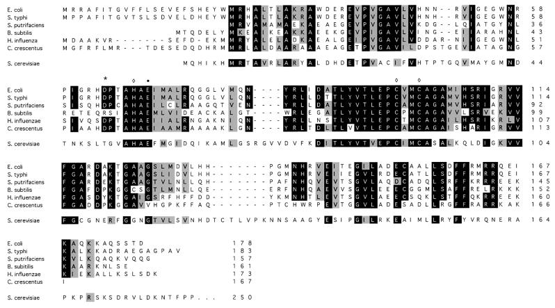
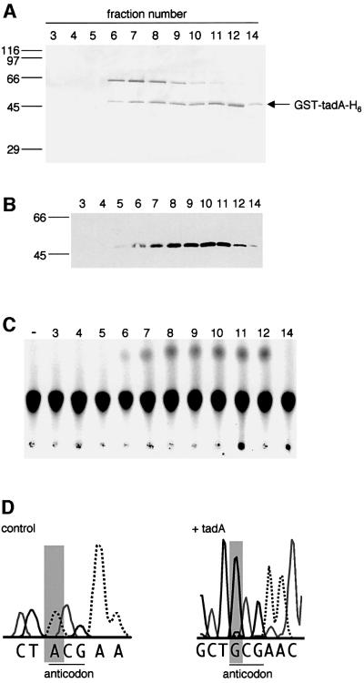
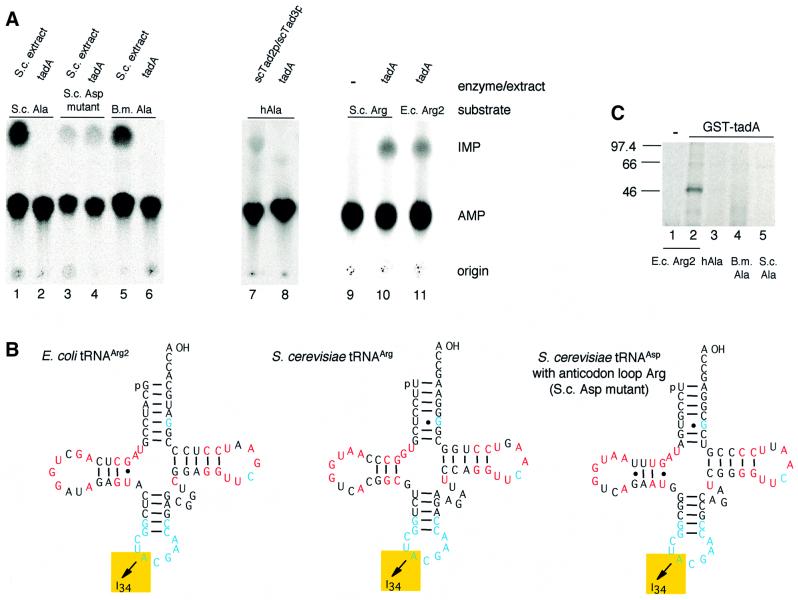
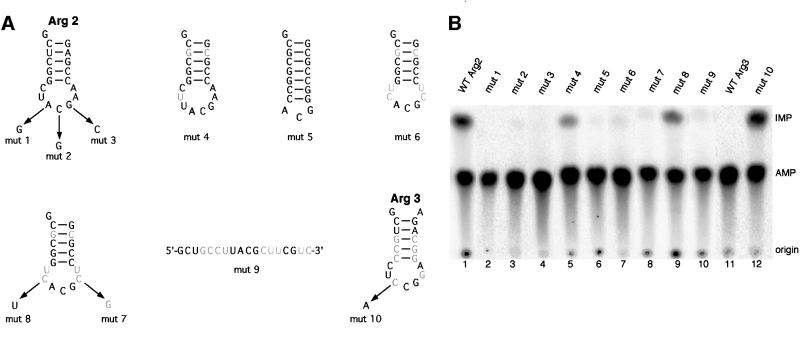
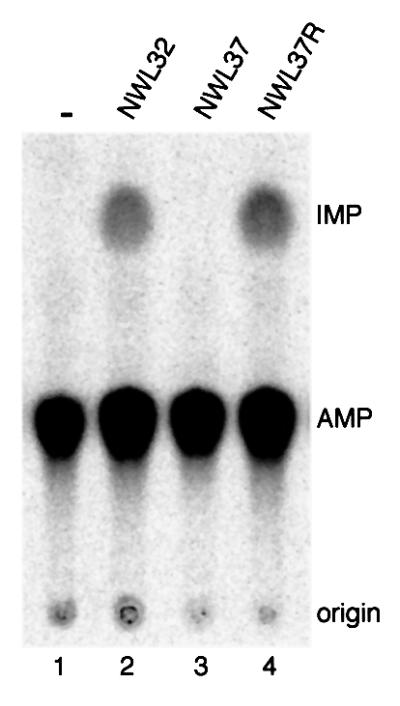
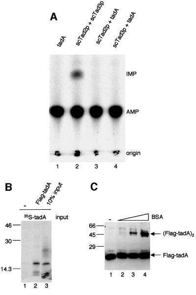
We report the characterization of tadA, the first prokaryotic RNA editing enzyme to be identified. Escherichia coli tadA displays sequence similarity to the yeast tRNA deaminase subunit Tad2p. Recombinant tadA protein forms homodimers and is sufficient for site-specific inosine formation at the wobble position (position 34) of tRNA(Arg2), the only tRNA having this modification in prokaryotes. With the exception of yeast tRNA(Arg), no other eukaryotic tRNA substrates were found to be modified by tadA. How ever, an artificial yeast tRNA(Asp), which carries the anticodon loop of yeast tRNA(Arg), is bound and modified by tadA. Moreover, a tRNA(Arg2) minisubstrate containing the anticodon stem and loop is sufficient for specific deamination by tadA. We show that nucleotides at positions 33-36 are sufficient for inosine formation in mutant Arg2 minisubstrates. The anticodon is thus a major determinant for tadA substrate specificity. Finally, we show that tadA is an essential gene in E.coli, underscoring the critical function of inosine at the wobble position in prokaryotes.


Figures

None
Fig. 1. Bacterial genomes encode a protein (tadA) related to Tad2p of S.cerevisiae. Multiple sequence alignment of E.coli tadA, yeast Tad2p and putative tadA sequences from different bacteria. Residues conserved in >83% of the proteins are shown in black, similar amino acids in gray. The three putative Zn2+-chelating residues (diamond) and the glutamate thought to mediate proton transfer (bullet) are marked. The position of the point mutation (D64E) in the tadA mutant NWL37 is indicated by an asterisk. The alignment was generated with the Clustal_W software at the European Bioinformatics Institute (Thompson et al., 1994).
None
Fig. 2. tadA specifically deaminates A34 in tRNAArg2. SDS– polyacrylamide gel stained with silver (A), western blot (B) and tRNA editing assay (C) of the final gel filtration column of the purification of recombinant tadA. A 20 µl aliquot of each fraction was separated on a 12% SDS–polyacrylamide gel (A) and transferred to a nitrocellulose membrane for detection with antibodies (B). The western blot was probed with a mouse α-GST monoclonal antibody. Fraction numbers are indicated at the top, the molecular masses of the size standards in kDa on the left. (C) Two microliters of the fractions indicated at the top were incubated with [33P]ATP-labeled tRNAArg2 for 30 min at 37°C. Reactions were treated with P1 nuclease and the products separated by one-dimensional TLC. The positions of AMP, IMP and the origin are indicated on the right. The position of IMP was verified with unlabeled 5′ IMP. In lane ‘–’, tRNAArg2 was incubated with buffer only. (D) Sequence analysis of in vitro modified tRNAArg2. The nucleotide sequence surrounding the anticodon is shown. tRNA from reactions shown in (C) was amplified by RT–PCR and sequenced. The nucleotide at position 34 is shaded gray, the anticodon is underlined. Control: tRNAArg2 incubated with buffer only.
None
Fig. 3. Substrate specificity of tadA. (A) tRNA-editing assay with tadA and different tRNAs. tRNA substrates were incubated with either 2 ng of recombinant His6-tadA (lanes 2, 4, 6, 8, 10 and 11), 20 ng of recombinant scTad2p/scTad3p (lane 7) or 40 µg of S.cerevisiae total protein (lanes 1, 3 and 5). One hundred femtomoles of tRNAAla from S.cerevisiae (lanes 1 and 2), a tRNAAsp mutant from S.cerevisiae (lanes 3 and 4), tRNAAla from B.mori (lanes 5 and 6), human tRNAAla (lanes 7 and 8), S.cerevisiae tRNAArg (lanes 9 and 10) and tRNAArg2 from E.coli (lane 11) were used. Mock incubation with WT yeast tRNAAsp did not result in I formation (result not shown). All reactions were incubated for 1 h at 30°C and processed as described in the legend to Figure 2 (see also Materials and methods). (B) Cloverleaf structure of E.coli tRNAArg2, S.cerevisiae tRNAArg and S.cerevisiae tRNAAsp with the anticodon loop of tRNAArg. Completely conserved nucleotides and nucleotides conserved as purines or pyrimidines between tRNAs from different species are shown in red (Klingler and Brutlag, 1993). Nucleotides that are conserved in addition between the three tRNAs are depicted in blue. (C) UV-cross-linking experiments of recombinant tadA and different tRNAs. A 400 ng aliquot of GST–tadA (lanes 2–5) and 100 fmol of [33P]ATP-labeled tRNA were irradiated and samples treated with RNaseA. Proteins were separated on a 12% SDS–polyacrylamide gel and gels exposed to a phosphoimager screen. E.c., E.coli; S.c., S.cerevisiae; B.m., B.mori; Arg2, tRNAArg2; hAla, human tRNAAla; B.m. Ala, B.mori tRNAAla; S.c. Ala, S.cerevisiae tRNAAla.
None
Fig. 4. RNA minisubstrates derived from E.coli tRNAArg2 and tRNAArg3 are sufficient for deamination by tadA in vitro. (A) Schematic drawing of Arg minisubstrates that were tested in vitro. Nucleotides shown in gray are mutated compared with WT Arg2. A at the wobble position of the anticodon is shown in bold. Mutations in each minisubstrate are indicated by arrows. (B) Editing assay with minisubstrates and tadA. A 25–100 fmol aliquot of minisubstrate, 100 ng of recombinant Flag-tadA-His6 and 500 ng of BSA were incubated in a total volume of 25 µl. WT Arg2 (lane 1), mutant 1 (lane 2), mutant 2 (lane 3), mutant 3 (lane 4), mutant 4 (lane 5), mutant 5 (lane 6), mutant 6 (lane 7), mutant 7 (lane 8), mutant 8 (lane 9), mutant 9 (lane 10), WT Arg3 (lane 11) and mutant 10 (lane 12) were used. All reactions were incubated for 45 min at room temperature and processed as described in the legend to Figure 2 (see also Materials and methods).
None
Fig. 5. The point mutation D64E abolishes tadA activity in vitro. Lane 1, no protein; lanes 2–4, 40 µg of total protein of E.coli extract. All reactions were incubated for 2 h at 37°C and processed as described in the legend to Figure 2 (see also Materials and methods).
None
Fig. 6. tadA cannot replace yeast Tad2p in vitro and forms homodimers. (A) Recombinant yeast Tad2p, Tad3p and E.coli tadA were pre-incubated as indicated at the top and tested for activity in vitro with tRNAAla from B.mori. As a control, the yeast Tad2p/Tad3p complex was used (lane 2). (B) Flag pull-down assay with in vitro translated tadA. 35S-labeled tadA was incubated with Flag-tagged tadA (lane 2) or buffer (lane 1) and subsequently bound to Flag–agarose and eluted. Ten percent of the input is shown in lane 3. Proteins were separated on a 12% SDS–polyacrylamide gel. Molecular masses of the protein standards are indicated in kDa on the left. (C) Chemical cross-linking of recombinant tadA. A 300 ng aliquot of tadA (lanes 2–4) was incubated in a reaction mixture containing tris(2,2′-bipyridyl) dichlororuthenium(II)hexahydrate, APS and BSA (90 ng, 0.9 µg and 9 µg, respectively). tadA incubated with buffer only is shown in lane 1. Reaction products were separated on a 12% SDS–polyacrylamide gel and blotted onto a nitrocellulose membrane. tadA was detected with a mouse anti-Flag antibody. Molecular masses of protein standards are indicated in kDa on the left.
None
Fig. 7. tadA is an essential gene. (A) The sacB counterselection procedure. The wild-type chromosomal copy of tadA was exchanged via a two-step homologous recombination reaction with a gene copy disrupted by the insertion of a kanamycin-resistance cassette. After the second recombination step, colonies were analyzed by their antibiotic-resistance profile for the presence or absence of the Kan cassette in the tadA locus and for mutational inactivation of the sacB gene. The mutation in sacB is indicated by an asterisk. (B) Summary of the clones obtained by the strategy described in (A). The total number of sucrose-resistant colonies analyzed is indicated in the first column (Total clones). The number of isolated colonies with a disrupted tadAtadA) gene in the chromosome is shown in the second column and the number of colonies with a restored WT tadA locus (WT) after the second recombination step is shown in the third column. Colonies that had acquired sucrose resistance through sacB inactivation are listed in the fourth column (sacB). Plasmids pJW30 and pJW117 carry disrupted and WT copies of the tadA ORF, respectively. (C) PCR analysis of WT (lane 1) and ΔtadA (lane 2) genomic E.coli DNA. Primers are indicated by arrows and anneal 500 bp upstream of the tadA ORF and at the 3′ end of the ORF, respectively. Sizes of standards (lane M) are indicated in bp on the left.

Similar articles

Cited by

References

    1. Altman S. and Kirsebom,L. (1999) Ribonuclease P. In Gesteland,R.F., Cech,T.R. and Atkins,J.F. (eds), The RNA World. Cold Spring Harbor Laboratory Press, Cold Spring Harbor, NY.
    1. Auxilien S., Crain,P.F., Trewyn,R.W. and Grosjean,H. (1996) Mechanism, specificity and general properties of the yeast enzyme catalysing the formation of inosine 34 in the anticodon of transfer RNA. J. Mol. Biol., 262, 437–458. - PubMed
    1. Basilio C., Wahba,A.J., Lengyel,P., Speyer,J.F. and Ochoa,S. (1962) Synthetic polynucleotides and the amino acid code. Proc. Natl Acad. Sci. USA, 48, 613–616. - PMC - PubMed
    1. Bass B.L. (1997) RNA editing and hypermutation by adenosine deamination. Trends Biochem. Sci., 22, 157–162. - PubMed
    1. Bass B.L., Nishikura,K., Keller,W., Seeburg,P.H., Emeson,R.B., O’Connell,M.A., Samuel,C.E. and Herbert,A. (1997) A standardized nomenclature for adenosine deaminases that act on RNA. RNA, 3, 947–949. - PMC - PubMed

Publication types