. 2002 Jul 15;21(14):3863-72.
doi: 10.1093/emboj/cdf383.

Crystal structure of human 53BP1 BRCT domains bound to p53 tumour suppressor

Affiliations

Crystal structure of human 53BP1 BRCT domains bound to p53 tumour suppressor

Dean J Derbyshire et al. EMBO J. .

Erratum in

  • EMBO J 2002 Nov 1;21(21):5953

Abstract

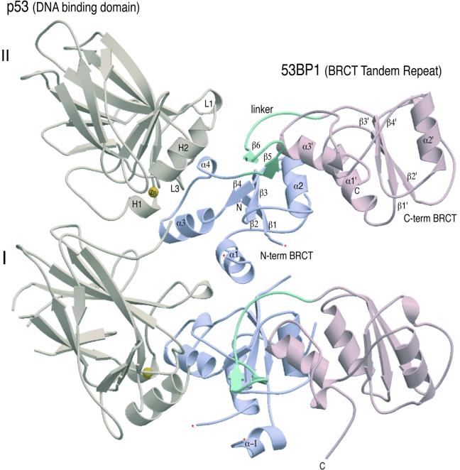
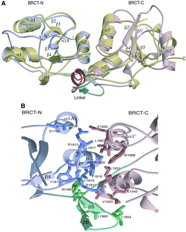
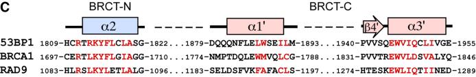
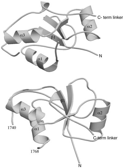
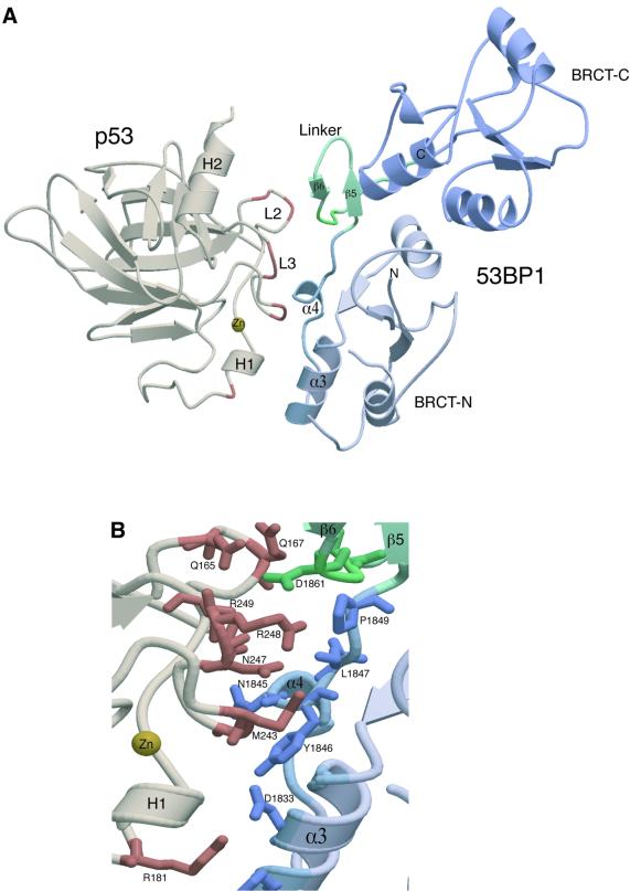
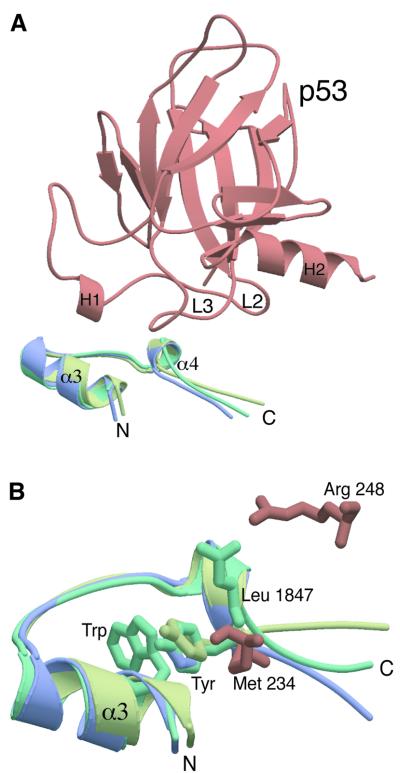
The BRCT (BRCA1 C-terminus) is an evolutionary conserved protein-protein interacting module found as single, tandem or multiple repeats in a diverse range of proteins known to play roles in the DNA-damage response. The BRCT domains of 53BP1 bind to the tumour suppressor p53. To investigate the nature of this interaction, we have determined the crystal structure of the 53BP1 BRCT tandem repeat in complex with the DNA-binding domain of p53. The structure of the 53BP1-p53 complex shows that the BRCT tandem repeats pack together through a conserved interface that also involves the inter-domain linker. A comparison of the structure of the BRCT region of 53BP1 with the BRCA1 BRCT tandem repeat reveals that the interdomain interface and linker regions are remarkably well conserved. 53BP1 binds to p53 through contacts with the N-terminal BRCT repeat and the inter-BRCT linker. The p53 residues involved in this binding are mutated in cancer and are also important for DNA binding. We propose that BRCT domains bind to cellular target proteins through a conserved structural element termed the 'BRCT recognition motif'.


Figures

None
Fig. 1. Crystal structure of the 53BP1–p53 complex. Structure of the asymmetric unit containing four molecules, two heterodimers (I and II) of p53 DNA-binding domain (brown) in complex with the BRCT tandem repeat of 53BP1 (blue and pink). The heterodimers pack together mainly through contacts made between the N-terminal region of 53BP1. There are no contacts made between the adjacent p53 molecules. The single zinc atom bound to p53 is shown in yellow and the 53BP1 inter-domain linker is shown in green. ‘Missing’ residues in the structure are denoted by an asterisk.
None
Fig. 2. Structural features of the 53BP1 tandem BRCT repeat. (A) A secondary structure representation of the superimposition of the BRCT tandem repeats from human BRCA1 and 53BP1. The N- and C-terminal BRCT domains of 53BP1 are coloured in light blue and pink, respectively, whilst both BRCT domains of human BRCA1 are coloured gold. The linker regions of each tandem repeat are also highlighted; BRCA1 (a loop–helix–loop structure) in orange and 53BP1 (a β-ribbon-like motif) in green. ‘Missing’ residues in the structure are denoted by an asterisk. (B) Intra-molecular interactions at the inter-BRCT repeat interface of 53BP1. The side chains from the first BRCT repeat are shown in blue, those from the linker are in green and those from the second BRCT repeat are pink. The α2 helix of the first repeat together with α1′ and α3′ helices of the second repeat form a three-helical bundle that is stabilized by α1A and the β-hairpin linker (β5–β6). (C) An amino acid sequence alignment of the regions of BRCA1, 53BP1 and RAD9 that are predicted to form BRCT–BRCT interfaces. Residues that constitute this interface in human 53BP1, as well as conserved residues in human BRCA1 and Sacchromyces cerevisiae RAD9, are coloured red.
None
Fig. 3. Contacts between 53BP1 BRCT–BRCT domains. The N-terminal BRCT domains form non-crystallographic interactions. The interface is composed of non-specific interactions between residues in α1 and an extended loop region, shown in shaded colour.
None
Fig. 4. Structure of the 53BP1–p53 interface. (A) The heterodimeric complex of 53BP bound to p53. The p53 binding region of 53BP1 forms an extended structure from the N-terminal region of the first BRCT (shown in light blue) to the central helical region of the inter-domain linker (α4). The 53BP1 contacts (shown in brown) are with highly conserved residues on the L2 and L3 loops of p53. The L2 and L3 loops form rigid hairpin structures that are stabilized by a single co-ordinating zinc atom (yellow). (B) Specific amino acid contacts at the complex interface between 53BP1 (blue) and p53 (brown). The inter-domain β-turn (β5-β6) is shown in green.
None
Fig. 5. A conserved protein binding motif in BRCT domains. (A) Superimposition of the conserved p53-binding motif. This shows a superimposition of the p53-binding interface of 53BP1 (green; residues 1829–1849) with the comparable regions of the BRCT domains of BRCA1 (blue; 1716–1738) and XRCC1 (yellow; 72–94). The Cα backbone of these regions overlap with an r.m.s.d. of <1 Å. The position of this motif in relation to p53 binding is also shown (p53 core domain is shown in brown). (B) The highly conserved Trp residue on α3 is positioned in an identical position in all three structures (for clarity only a single Trp is shown) and stacks against the conserved Tyr residue (the superimposed Tyr shown). Two conserved 53BP1–p53 interactions are shown, Arg 248–Leu 1847 and Met 243–Tyr1846.

References

    1. Anderson L., Henderson,C. and Adachi,Y. (2001) Phosphorylation and rapid relocalization of 53BP1 to nuclear foci upon DNA damage. Mol. Cell. Biol., 21, 1719–1729. - PMC - PubMed
    1. Bargonetti J., Manfredi,J.J., Chen,X., Marshak,D.R. and Prives,C. (1993) A proteolytic fragment from the central region of p53 has marked sequence-specific DNA-binding activity when generated from wild-type but not from oncogenic mutant p53 protein. Genes Dev., 7, 2565–2574. - PubMed
    1. Bork P., Hofmann,K., Bucher,P., Neuwald,A.F., Altschul,S.F. and Koonin,E.V. (1997) A superfamily of conserved domains in DNA damage-responsive cell cycle checkpoint proteins. FASEB J., 11, 68–76. - PubMed
    1. Brünger A.T. et al. (1998) Crystallography and NMR system: a new software suite for macromolecular structure determination. Acta Crystallogr. D Biol. Crystallogr., 54, 905–921. - PubMed
    1. Callebaut I. and Mornon,J.P. (1997) From BRCA1 to RAP1: a widespread BRCT module closely associated with DNA repair. FEBS Lett., 400, 25–30. - PubMed

Publication types

MeSH terms

Substances