. 2002 Jul 15;21(14):3888-97.
doi: 10.1093/emboj/cdf379.

Accessory factors determine the order of strand exchange in Xer recombination at psi

Affiliations

Accessory factors determine the order of strand exchange in Xer recombination at psi

Migena Bregu et al. EMBO J. .

Abstract

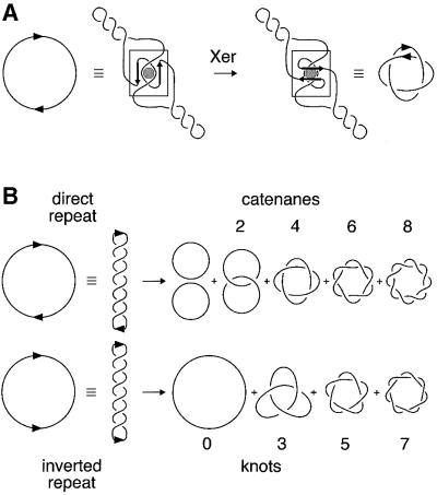
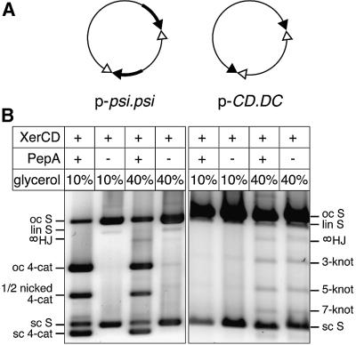
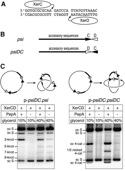
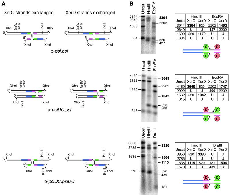
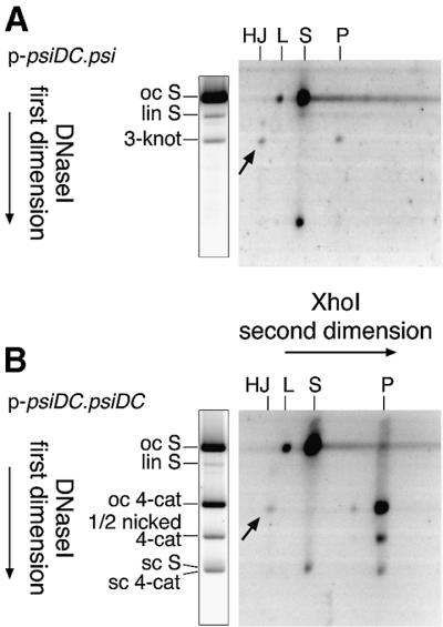
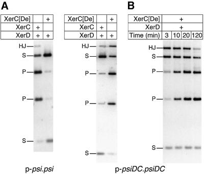
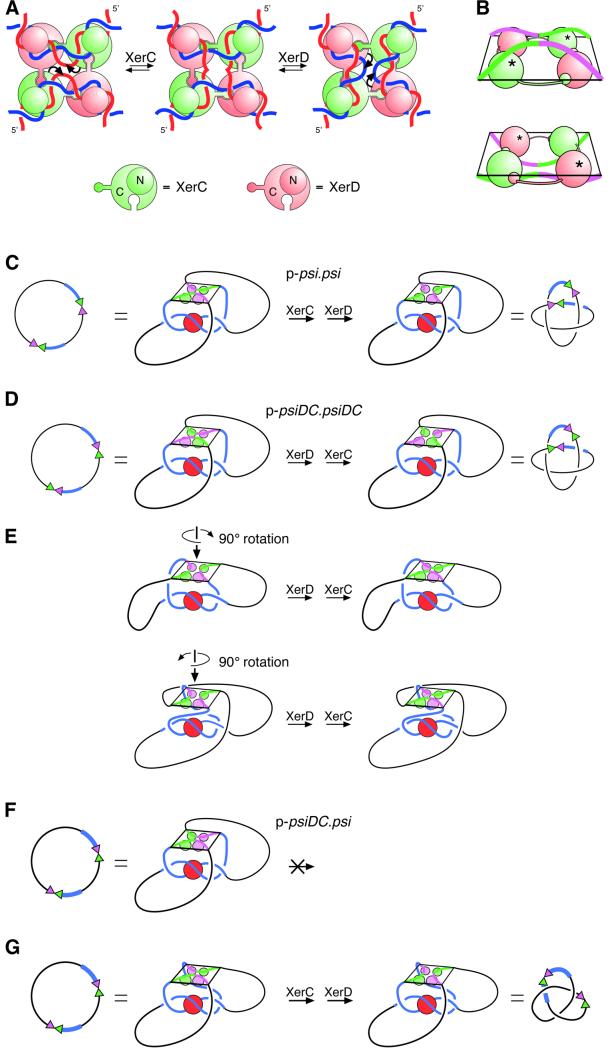
Xer site-specific recombination in Escherichia coli converts plasmid multimers to monomers, thereby ensuring their correct segregation at cell division. Xer recombination at the psi site of plasmid pSC101 is preferentially intramolecular, giving products of a single topology. This intramolecular selectivity is imposed by accessory proteins, which bind at psi accessory sequences and activate Xer recombination at the psi core. Strand exchange proceeds sequentially within the psi core; XerC first exchanges top strands to produce Holliday junctions, then XerD exchanges bottom strands to give final products. In this study, recombination was analysed at sites in which the psi core was inverted with respect to the accessory sequences. A plasmid containing two inverted-core psi sites recombined with a reversed order of strand exchange, but with unchanged product topology. Thus the architecture of the synapse, formed by accessory proteins binding to accessory sequences, determines the order of strand exchange at psi. This finding has important implications for the way in which accessory proteins interact with the recombinases.


Figures

None
Fig. 1. Topology of site-specific recombination reactions. (A) Xer recombination at psi occurs only after the formation of a synaptic complex with a defined local structure (shown boxed). This productive synapse, formed by wrapping the accessory sequences of two recombination sites around the accessory protein PepA, traps a specific number of topological nodes. Strand exchange occurs by a defined mechanism and the product is a right-handed four-noded catenane with antiparallel psi sites. (B) Many site-specific recombination systems display no topological specificity. A random number of supercoils are trapped when the recombination sites come together, and recombination generates products of mixed topology. Sites in direct repeat yield two circles, which are either unlinked, or linked in catenanes with even numbers of topological crossings or nodes, as shown. Sites in an inverted repeat yield single circular products, which are either unknotted or knotted with an odd number of nodes. Knots and catenanes produced in these reactions are generally members of the torus family of knots and catenanes, as shown.
None
Fig. 2. Recombination at psi sites with and without accessory sequences. (A) Maps of p-psi.psi, containing two psi sites in direct repeat, and p-CD.DC, containing two psi cores without accessory sequences in inverted repeat. Accessory sequences are shown as thick black lines; XerC- and XerD-binding sites in the psi core are shown as filled and open triangles, respectively. (B) Topological analysis of recombination products. Plasmids were reacted with XerC and XerD with or without PepA, with either 10 or 40% glycerol as indicated. Reactions were nicked and run on a 0.7% agarose gel. Bands are indicated as follows: oc S, open circle substrate; lin S, linear substrate; sc S, supercoiled substrate; oc 4-cat, four-noded catenated product nicked on both circles; 1/2 nicked 4-cat, four-noded catenated product nicked on the large circle but still supercoiled on the small circle; sc 4-cat, fully supercoiled four-noded catenane; 3-knot, 5-knot and 7-knot, nicked knotted inversion products with three, five and seven nodes, respectively; ∞HJ, HJ intermediate nicked at the HJ on the recombinant strand, with consequent loss of any knotting or catenation.
None
Fig. 3. Recombination at psiDC. (A) Sequence of the psi core showing XerC- and XerD-binding and cleavage sites. (B) Diagram of the psi and psiDC sites, showing accessory sequences, to which PepA binds, and the core site with XerC- and XerD-binding sites represented as filled and open triangles, respectively. (C) Diagrams of p-psiDC.psi and p-psiDC.psiDC and their major recombination products. Plasmids were reacted with XerC and XerD in the presence or absence of PepA, with 10 or 40% glycerol (as indicated). Reactions were nicked and run on a 0.7% agarose gel. Bands are indicated as in Figure 2.
None
Fig. 4. Analysis of HJ intermediates formed by recombination at psi and psiDC. (A) HJ intermediates made by XerC or XerD strand exchange can be distinguished by the recombinant strands they contain. XhoI-cleaved χ-form HJs, 3′ end-labelled and then cleaved with restriction enzymes give different patterns of single-stranded DNA fragment depending on which strands have been exchanged. Accessory sequences are shown in blue, XerC- and XerD-binding sites are shown in green and pink, respectively, and 3′ end labels are indicated by asterisks. (BXhoI-cleaved HJs from p-psi.psi, p-psiDC.psi and p-psiDC.psiDC were purified and 3′ end-labelled with 32P. Labelled χ DNA was run on a strand-separating agarose gel uncut or cleaved with HindIII, EcoRV or DraIII. Predicted sizes of labelled single-stranded DNA fragments for χ-forms produced by XerC or XerD strand exchange before or after cleavage with the appropriate restriction enzymes are tabulated. U indicates fragments unchanged by the restriction enzyme. Fragment sizes diagnostic for XerC or XerD strand exchanges are shown in bold. Sites taking part in each reaction are shown diagrammatically below each table. Curly arrows indicate recombinase partners initiating the first strand exchange reaction.
None
Fig. 5. Two-dimensional gel analysis of HJs formed by recombination between psi and psiDC sites. (A) Analysis of products formed by recombination on p-psiDC.psi. (B) Analysis of products formed by recombination on p-psiDC.psiDC. The first dimension separated substrates and products according to their topology, and the second dimension separated products according to their restriction pattern. Arrows indicate the positions of HJ intermediates on each gel. Migration of three-noded knots, four-noded catenanes and other species in the first dimension is indicated to the left of a duplicate first- dimension lane as in the legend to Figure 2. Second-dimension mobilities are shown above the gels as HJ, HJ intermediates; L, linear substrate; S, substrate restriction fragment; and P, product restriction fragment. Small product and substrate restriction fragments are not shown. Partial digestion with XhoI gave some linear substrate at the oc S position, and some uncut (open circle) large product circle at the four-node position in (B).
None
Fig. 6. Xer recombination at psi and psiDC in the presence of XerC[De]. (A) Recombination of p-psi.psi and p-psiDC.psiDC in the presence of XerD and either XerC[De] or XerC. (B) Time course of recombination on p-psiDC.psiDC with XerC[De] and XerD. Reactions contained 40% glycerol and were cleaved with XhoI. Bands are indicated as follows: HJ, HJ intermediates; S, substrate fragments; P, product fragments.
None
Fig. 7. Models for recombination at psi and psiDC. (A) The proposed mechanism of strand exchange by XerC and XerD at the psi core, based on X-ray crystal structures of the Cre recombinase bound to its recombination site. XerC (green) and XerD (pink) bind to a pair of psi core sites in an antiparallel conformation. A C-terminal extension on each monomer binds to the C-terminal domain of one of its neighbours in a cyclical arrangement. All four monomers bind to the DNA with their N-terminal domains (shown as small circles above the DNA) on one side of the HJ and their C-terminal domains (shown as large circles below the DNA) on the other side. XerC catalyses cleavage, strand exchange and re-ligation of one pair of strands (denoted by curly arrows) to form a HJ intermediate. XerD then exchanges the other pair of strands to form the recombinant product. Recombination can also occur by the reverse of this process (right to left) with XerD strand exchange preceding XerC strand exchange. (B) Perspective view of core–recombinase complexes in which XerC (top panel; equivalent to the left panel in A) or XerD (bottom panel; equivalent to the right panel in A) is about to catalyse strand exchange to form a HJ. The diagrams have been aligned so that DNA connectivity and the topological outcome of a complete strand exchange reaction will be the same in both cases. In this view, C-terminal domains have to be below the DNA for XerC strand exchange to proceed first, and above the DNA for XerD strand exchange to proceed first. XerC and XerD C-terminal domains are shown as green and pink spheres, respectively. N-terminal domains have been omitted for clarity. Active recombinase monomers are indicated with asterisks. (C) Synapsis of psi sites and interwrapping of accessory sequences around PepA define the order of strand exchange by XerC and XerD. During recombination between two psi sites, the accessory sequences (blue) of two psi sites wrap around PepA, with the psi cores brought together in an antiparallel fashion. XerC protein and binding sites are shown in green, and XerD protein and binding sites are shown in pink. This synapse is equivalent to that shown in Figure 1A. The recombinase–core complex is constrained so that the C-termini face towards PepA and the accessory sequences. By comparison with (A), it can be seen that the psi cores have been brought together such that XerC strand exchange will precede XerD strand exchange. The product is a four-noded catenane. (D) Two psiDC sites can form a synapse in which the DNA follows exactly the same path as in (C). In this complex, XerC occupies the position normally occupied by XerD, and vice versa. XerD strand exchange proceeds first in this synapse, and the product is a four-noded catenane. (E) The synapse shown in (D) can be rearranged into two geometrically different, but topologically equivalent synapses, by rotating the recombinase–core complex by ±90° about its 2-fold symmetry axis. XerC and XerD occupy the same positions in this synapse as in the synapse between wild-type psi sites, but the DNA path is altered to accommodate the psiDC sites. XerD strand exchange will proceed first and the product will be the observed four-noded catenane in both cases. (F) On p-psiDC.psi, interwrapping of accessory sequences as in (A) would bring the two cores together in a parallel conformation with forbidden XerC–XerD interactions. (G) To bring the two sites together in an antiparallel fashion, one of the cores must be rotated through 180° relative to its position in (F). If the core of psiDC is rotated, as depicted, XerC strand exchange will proceed first and the product will be a three-noded knot, as observed. However, an equivalent rotation of the psi core would give the same three-noded product but XerD strand exchange would proceed first. Alternatively, both cores might be rearranged to bring about the required antiparallel alignment.

References

    1. Alén C., Sherratt,D.J. and Colloms,S.D. (1997) Direct interaction of aminopeptidase A with recombination site DNA in Xer site-specific recombination. EMBO J., 16, 5188–5197. - PMC - PubMed
    1. Arciszewska L., Grainge,I. and Sherratt,D. (1995) Effects of Holliday junction position on Xer-mediated recombination in vitro. EMBO J., 14, 2651–2660. - PMC - PubMed
    1. Arciszewska L.K., Grainge,I. and Sherratt,D.J. (1997) Action of site-specific recombinases XerC and XerD on tethered Holliday junctions. EMBO J., 16, 3731–3743. - PMC - PubMed
    1. Arciszewska L.K., Baker,R.A., Hallet,B. and Sherratt,D.J. (2000) Coordinated control of XerC and XerD catalytic activities during Holliday junction resolution. J. Mol. Biol., 299, 391–403. - PubMed
    1. Aussel L., Barre,F.-X., Aroyo,M., Stasiak,A., Stasiak,A.Z. and Sherratt,D.J. (2002) FtsK is a DNA motor protein that activates chromosome dimer resolution by switching the catalytic state of XerC and XerD recombinases. Cell, 108, 195–205. - PubMed

Publication types