. 2002 Jul 15;21(14):3898-907.
doi: 10.1093/emboj/cdf369.

Replication fork collapse at replication terminator sequences

Affiliations

Replication fork collapse at replication terminator sequences

Vladimir Bidnenko et al. EMBO J. .

Abstract

Replication fork arrest is a source of genome re arrangements, and the recombinogenic properties of blocked forks are likely to depend on the cause of blockage. Here we study the fate of replication forks blocked at natural replication arrest sites. For this purpose, Escherichia coli replication terminator sequences Ter were placed at ectopic positions on the bacterial chromosome. The resulting strain requires recombinational repair for viability, but replication forks blocked at Ter are not broken. Linear DNA molecules are formed upon arrival of a second round of replication forks that copy the DNA strands of the first blocked forks to the end. A model that accounts for the requirement for homologous recombination for viability in spite of the lack of chromosome breakage is proposed. This work shows that natural and accidental replication arrests sites are processed differently.


Figures

None
Fig. 1. Schematic representation of Lac-Phe-Ter chromosome. (A) Chromosome on which replication is blocked at Terphe and Terlac. (B) Cleavage of both leading and lagging strands. (C) Cleavage of two strands of the same polarity (leading or lagging strands), at both Ter sites. (D) DNA replication initiated at oriC and progressing to Terphe and Terlac. Natural terminators are symbolized by gray indented rectangles, and Terphe and Terlac by white indented rectangles. The chromosome origins (oriC) are shown as small circles. Each line represents double-stranded DNA. Cleavage sites are indicated by black arrows.
None
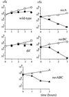
Fig. 2. recA, recB and ruvABC mutations are lethal in the Lac-Phe-Ter strain, dif deletion is not. At time 0, a culture growing in LB supplemented with glucose was centrifuged, resuspended in LB medium and split into two parts to which either glucose or arabinose was added. At each indicated time, an aliquot was withdrawn and appropriate dilutions were plated on LB medium supplemented with glucose. Open symbols, growth in glucose; closed symbols, growth in arabinose. Circles, Lac-Phe-Ter strain; squares, Lac-Phe-Ter dif; rectangles, Lac-Phe-Ter recA; diamonds, Lac-Phe-Ter recBC; triangles, Lac-Phe-Ter ruvABC.
None
Fig. 3. Tus induction leads to the appearance of a linear fragment upstream of Terphe only. Lanes 1 and 2, ethidium bromide staining of a pulsed-field gel of a NotI-digested Lac-Phe-Ter recB chromosome. Lanes 3 and 4, Southern blot of the same gel using recN DNA as a probe. Lanes 5 and 6, Southern blot of the same gel using recO DNA as a probe. Lanes 1, 3 and show 5 growth in glucose (G), and lanes 2, 4 and 6 show growth in arabinose (A). Sizes of the AB1157 chromosome NotI fragments are indicated on the left (Perkins et al., 1993). Because of replication arrest at Terphe and Terlac, the intensity of DNA fragments located downstream of these Ter sites is decreased in arabinose compared with glucose. It can be noted that the smallest Not band that hybridizes with recN is smeary, probably due to the in vivo erosion of the DNA end by various single-stranded exonucleases, such as SbcB, RecJ or ExoVII. The position of the gel origin (O), linear NotI fragments encompassing Terphe (L), replication intermediate molecules formed upon fork blockage at Terphe (Y) and the expected positions of migration of linear fragments from the upstream NotI to Terphe (NotI–Ter) and from Terphe to the downstream NotI site (TerNotI) are indicated. A schematic representation of the NotI fragment encompassing the Terphe site is shown. The positions of recN and recO genes used as probes and of Terphe are indicated. The indented rectangle represents a Tus-bound Terphe site. Chromosome replication proceeds from left to right. The distances from Terphe to upstream and downstream NotI sites are indicated (34.7 and 222.9 kb, respectively).
None
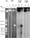
Fig. 4. Tus induction leads to the formation of 2 Mb linear molecules that hybridize with the oriC half of the chromosome only. Lane 1, Hanselina wingei (Hw) chromosome size marker. Lanes 2–4, ethidium bromide staining of a pulsed-field gel of intact recB (lane 2) or Lac-Phe-Ter recB (lanes 3–4) chromosome. Lanes 5 and 6, Southern blot of lanes 3 and 4 using the serA gene DNA as a probe (oriC-proximal probe). Lanes 7 and 8, Southern blot of the same lanes using the hisG gene DNA as a probe (terminal region probe). Lanes 3, 5 and 7 show growth in glucose, and lanes 2, 4, 6 and 8 show growth in arabinose. The position of the gel origin (O) is indicated. Schizosaccharomyces pombe chromosomes were also used routinely as size markers, indicating that linear chromosomes of 3–6 Mb migrate in pulsed-fields gels in the conditions used. It should be noted that for unknown reasons, the apparent intensities of bands in pulsed-field gels performed with entire E.coli chromosomes may be misleading, which is the reason why precise quantification of the amount of DNA in different regions of the gels was performed by counting [3H]thymidine-labeled DNA.
None
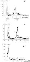
Fig. 5. Peak 2 is formed by newly synthesized DNA. The proportion of labeled DNA in each gel slice relative to the total amount of linear DNA migrating in a pulsed-field gel is shown. The gel origin is not shown. The positions of peak 1 and peak 2 are indicated. (A) Cells were grown for 3 h in [3H]thymidine glucose medium, washed and then shifted for 3 h to either [3H]thymidine glucose medium (dashed line, crosses) or to [3H]thymidine arabinose medium (full lines, squares). Peak 1 and peak 2 encompass all slices in which [3H]thymidine is significantly more abundant in glucose and in arabinose, respectively. (B) Formation of peak 2 during Tus induction. Cells were grown for 3 h in cold glucose medium, washed and then shifted to either glucose or arabinose tritiated medium. Dashed line with crosses, shift to [3H]thymidine glucose medium and incubation for 3 h. Full lines, shift to [3H]thymidine arabinose medium and incubation for: open squares, 1 h; gray squares, 2 h; filled squares, 3 h. (C) Peak 2 is not labeled when [3H]thymidine is present only prior to Tus induction. Cells were grown for 3 h in [3H]thymidine glucose medium, washed and then shifted to glucose or arabinose cold medium. Dashed line with crosses: shift to glucose cold LB and incubation for 3 h; full line with black squares: shift to arabinose cold LB and incubation for 3 h.
None
Fig. 6. Replication collapse model for the formation of Ter-induced linear DNA. In the first step, a clockwise (1C) and a counterclockwise (1CC) replication fork initiated at the chromosome origin (o) are blocked by Tus bound at Terphe and Terlac, respectively (indented rectangle). In the second step, a second round of replication has started from the origin (2C and 2CC). Black lines, template DNA; blue lines, strands of the first round of replication; red lines, strands of the second round of replication; yellow ovals, progressing replisomes. (A) Only the clockwise migrating forks are shown in this part of the model. (1) The second round of replication dislodges Tus. (2) The replisome is reassembled at the Tus-free LacTer site. (3) The linear DNA recombines with the homologous chromosome. A replisome is assembled at the recombination intermediates. (4) Resolution of Holliday junctions; the strands favored by migration of Holliday junctions toward the origin are exchanged. (B) Only the top chromatid with both clockwise and counterclockwise replication forks is shown here. Branch migration toward the origin, hence in two opposite directions, of Holliday junctions formed at clockwise and counterclockwise forks implies exchange of different strands at both junctions. (5) The first clockwise and counterclockwise forks reach the terminus. (6) The second replication forks reach the terminus region; a dimer molecule is formed. A chromosome dimer is also formed from the bottom chromatid (not shown), resulting in a cell that contains two dimeric chromosomes. (C) Only the clockwise migrating forks are shown here. (1) The second round of replication does not dislodge Tus and is blocked at Ter; linear DNA is formed by copy of the first strands to the end (collapse). (2) The linear DNA recombines with the homologous chromosome and a replisome is assembled at the recombination intermediates. (3) Holliday junctions are resolved; the exchanged strands are determined by the direction of branch migration, toward the origin. The forks reassembled at the recombination intermediates reach Ter. These forks dislodge Tus, and linear DNA is formed as the result of a second collapse reaction. (4) The linear DNA molecules recombine with the homologous chromosome and a replisome is assembled at the recombination intermediates. (5) Holliday junctions are resolved by RuvABC. Since productive branch migration is again directed toward the origin, the exchanged strands are the same as in the first homologous recombination reaction. (D) Only the top chromatid with both clockwise and counterclockwise replication forks is shown here. (6) The first clockwise and counterclockwise forks reach the terminus. (7) The second replication forks reach the terminus. Two monomers are formed from each chromatid, four per cell.

Similar articles

Cited by

References

    1. Aguilera A., Chavez,S. and Malagon,F. (2000) Mitotic recombination in yeast: elements controlling its incidence. Yeast, 16, 731–754. - PubMed
    1. Barre F.X., Soballe,B., Michel,B., Aroyo,M., Robertson,M. and Sherratt,D. (2001) Circles: the replication–recombination– chromosome segregation connection. Proc. Natl Acad. Sci. USA, 98, 8189–8195. - PMC - PubMed
    1. Bierne H., Ehrlich,S.D. and Michel,B. (1997a) Deletions at stalled replication forks occur by two different pathways. EMBO J., 16, 3332–3340. - PMC - PubMed
    1. Bierne H., Vilette,D., Ehrlich,S.D. and Michel,B. (1997b) Isolation of a dnaE mutation which enhances RecA-independent homologous recombination in the Escherichia coli chromosome. Mol. Microbiol., 24, 1225–1234. - PubMed
    1. Bussiere D.E. and Bastia,D. (1999) Termination of DNA replication of bacterial and plasmid chromosomes. Mol. Microbiol., 31, 1611–1618. - PubMed

Publication types