. 2002 Jul 23;99(15):10102-7.
doi: 10.1073/pnas.152152499. Epub 2002 Jul 10.

Global repression of exotoxin synthesis by staphylococcal superantigens

Affiliations

Global repression of exotoxin synthesis by staphylococcal superantigens

Nikola Vojtov et al. Proc Natl Acad Sci U S A. .

Abstract

Virulent Staphylococcus aureus strains typically produce and secrete large quantities of many extracellular proteins involved in pathogenesis. Such strains cause the classical staphylococcal lesion--local tissue destruction and aggressive inflammation accompanied by the massive influx of polymorphonuclear leukocytes, leading to the formation of pus. Most strains causing toxic shock syndrome, however, produce and secrete very small quantities of most exoproteins although they elaborate high levels of toxic shock syndrome toxin-1 (TSST-1). These strains cause local infections that are remarkably apurulent although potentially fatal owing to the superantigen. We have analyzed this disparity and have found that TSST-1 itself is a negative global regulator of exoprotein gene transcription. TSST-1 not only represses most exoprotein genes but determines its own high expression level by autorepression. We report also that a second superantigen, enterotoxin B, has similar regulatory properties.


Figures
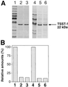
Figure 1
Figure 1
Effect of tst on exoprotein patterns. (A) Bacteria were grown to early stationary phase in CYGP in the absence of glucose, and supernatants were trichloroacetic acid-precipitated and analyzed by SDS/PAGE. Gels were stained with Coomassie brilliant blue and scanned. The following samples are shown: RN4282 with SaPI1 tsttetM insertional inactivation (RN6938) (lane 1), RN4282 tsttetM containing pRN5543∷tst(a.8) (pRN7119), expressing full-length tst (lane 2), RN4282 with SaPI1 carrying intact tst gene (lane 3), RN6734 (lane 4), RN6734 (SaPI1) strain (RN9131), expressing tst (lane 5), and RN6734 containing pRN5543∷tst(a.8) (pRN7119) and expressing tst (lane 6). Location of TSST-1 protein is indicated by the arrow. (B) Scanning data were used to compare the amounts of exoproteins produced by the various strains. Values for RN6938 (lane 1) and RN6734 (lane 4) were set to 100%. Amounts of exoproteins produced by derivative strains were normalized to this value, after subtracting the amount of TSST-1.
Figure 2
Figure 2
Mapping the locus of inhibition. At the top is a diagram of the SaPI1 tst locus, showing the promoter, the coding sequence (heavy black line), the inhibitory region (gray hatching), and the site of a frame-shift mutation. Deletions, introduced by the PCR, using primers listed in Table 2, are shown below, with the heavy black line indicating the extent of the remaining tst DNA. The effect of the progressive 3′ deletions on exoprotein expression is indicated by + for exoprotein inhibition or − for loss of the inhibitory activity. Numbers at the left, designating the individual deletions, are used throughout the text.
Figure 3
Figure 3
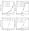
Effect of tst on exoprotein gene transcription. pRN5543 derivatives containing the intact tst gene (a.8) (pRN7119), a tst deletion, tst(a.5) (pRN7118), or tst with the frame shift mutation tst(FSh) (pRN7123) were transduced to separate RN6734 clones each with one of the following blaZ transcriptional fusions: (a) PsprblaZ (pRN7041) and (b) PlukSblaZ (pRN7040), with promoter-containing fragments of spr and lukS, respectively, or (c) Ptst(min)blaZ (with minimal promoter sequence of tst, defined as a +1 to −45 fragment, in respect to start of transcription). Expression of the reporter gene was monitored during growth at 37°C. (d) β-Lactamase expression kinetics for blaZ fusions with the intact tst, Ptst(a.8)∷blaZ (pRN7045), truncated tst, Ptst(a.5)∷blaZ (pRN7056), and frame-shifted tst, Ptst(FSh)∷blaZ (pRN7126).
Figure 4
Figure 4
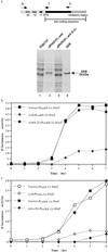
Effect of seb on expression of exoprotein genes. (a) Cultures of RN6734 containing the intact cloned seb, pRN5543∷seb (pRN7114) or a derivative with a large 3′ deletion, pRN5543∷seb(b.2) (pRN7116), were grown to early stationary phase, and the culture supernatants were analyzed for exoprotein patterns as in Fig. 1. Location of SEB protein is indicated by the arrow. (b) The plasmids used in a were introduced into a RN6734 derivative containing the Pseb(b.1)∷blaZ transcriptional fusion (pRN7112), and the kinetics of β-lactamase synthesis were monitored throughout growth. (c) pRN5543 derivatives containing either tst(a.8) (pRN7119) or intact seb (pRN7114) were introduced into RN6734 derivatives containing either Pseb(b.1)∷blaZ (pRN7112) or Ptst(a.1)∷blaZ (pRN7044), and β-lactamase activity was measured as in b.
Figure 5
Figure 5
Inhibition of inflammation by TSST-1-producing strain in vivo. Three mice were injected with either TSST-1-producing strain (RN4282) or isogenic tst strain (RN6938). Mice were inspected daily. Results shown are representative of each group.

Similar articles

Cited by

References

    1. McCormick J K, Yarwood J M, Schlievert P M. Annu Rev Microbiol. 2001;55:77–104. - PubMed
    1. Schlievert P, Osterholm M, Kelly J, Nishimura R. Ann Intern Med. 1982;96:937–940. - PubMed
    1. Todd J, Fishaut M, Kapral F, Welch T. Lancet. 1978;ii:1116–1118. - PubMed
    1. Hajjeh R A, Reingold A, Weil A, Shutt K, Schuchat A, Perkins B A. Emerg Infect Dis. 1999;5:807–810. - PMC - PubMed
    1. Bresler M J. West J Med. 1983;139:710–713. - PMC - PubMed

Publication types

LinkOut - more resources