

Different activation dynamics in multiple neural systems during simulated driving
- PMID: 12112769
- PMCID: PMC6872105
- DOI: 10.1002/hbm.10032
Different activation dynamics in multiple neural systems during simulated driving
Erratum in
- Hum Brain Mapp 2002 Oct;17(2):141-2
Abstract
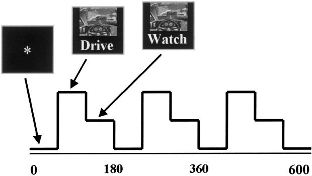
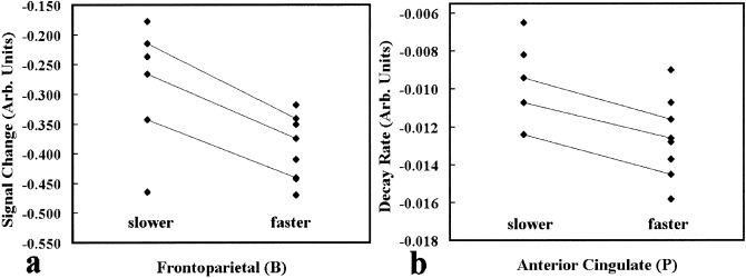
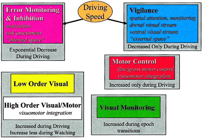
Driving is a complex behavior that recruits multiple cognitive elements. We report on an imaging study of simulated driving that reveals multiple neural systems, each of which have different activation dynamics. The neural correlates of driving behavior are identified with fMRI and their modulation with speed is investigated. We decompose the activation into interpretable pieces using a novel, generally applicable approach, based upon independent component analysis. Some regions turn on or off, others exhibit a gradual decay, and yet others turn on transiently when starting or stopping driving. Signal in the anterior cingulate cortex, an area often associated with error monitoring and inhibition, decreases exponentially with a rate proportional to driving speed, whereas decreases in frontoparietal regions, implicated in vigilance, correlate with speed. Increases in cerebellar and occipital areas, presumably related to complex visuomotor integration, are activated during driving but not associated with driving speed.
Copyright 2002 Wiley-Liss, Inc.
Figures

References
-
- Akshoomoff NA, Courchesne E, Townsend J (1997): Attention coordination and anticipatory control. Int Rev Neurobiol 41: 575–598. - PubMed
-
- Allen G, Buxton RB, Wong EC, Courchesne E (1997): Attentional activation of the cerebellum independent of motor involvement. Science 275: 1940–1943. - PubMed
-
- Ballard DH, Hayhoe MM, Salgian G, Shinoda H (2000): Spatio‐temporal organization of behavior. Spat Vis 13: 321–333. - PubMed
-
- Bell AJ, Sejnowski TJ (1995): An information maximization approach to blind separation and blind deconvolution. Neural Comput 7: 1129–1159. - PubMed
-
- Biswal BB, Ulmer JL (1999): Blind source separation of multiple signal sources of FMRI data sets using independent component analysis. J Comput Assist Tomogr 23: 265–271. - PubMed
Publication types
MeSH terms
Grants and funding
LinkOut - more resources
Full Text Sources
