

Capillary and cell wall permeability to potassium in isolated dog hearts
- PMID: 1211446
- PMCID: PMC2942799
- DOI: 10.1152/ajplegacy.1975.229.3.537
Capillary and cell wall permeability to potassium in isolated dog hearts
Abstract
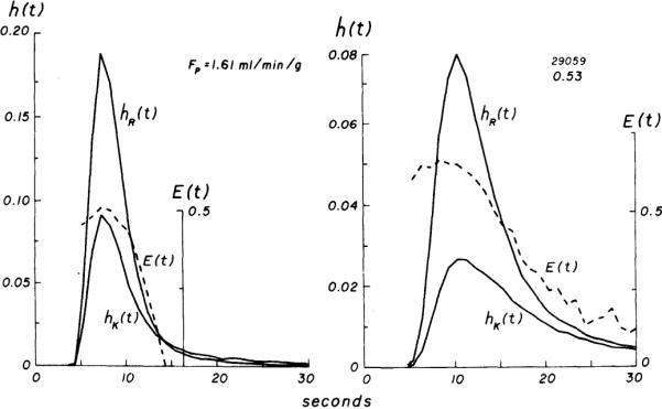
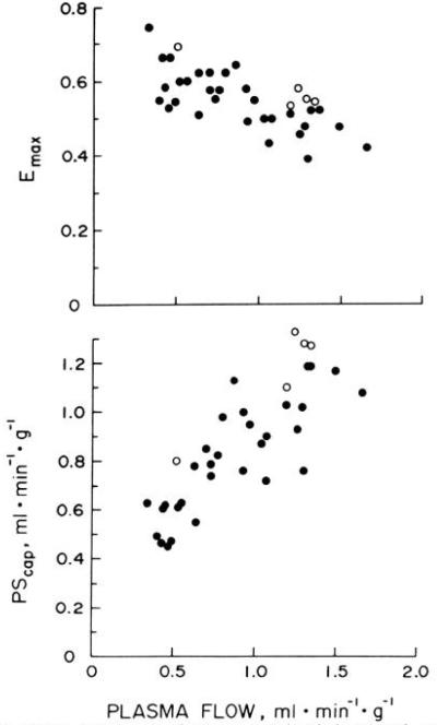
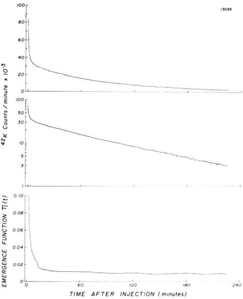
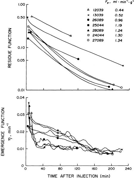
From venous tracer-dilution curves recorded after 36 pulse injections of 42KCl and 131I-labeled albumin into the coronary artery inflow of 15 isolated canine heart preparations, we calculated maximal fractional extractions (Emax) and capillary permeability-surface area products (PScap) for 42K+ over a range of plasma flows (FP) from 0.3 to 1.7 ml min-1 g-u. At low FP (less than 1.0), Emax was 0.60 +/- 0.0l (mean +/- SD) and PScap was 0.72 +/- 0.20 ml min-1 g-1; at high FP (greater than 1.0), Emax decreased to 0.49 +/- 0.05 and PScap increased to 1.06 +/- 0.18. Continuous recording (gamma detector) of residual myocardial 42K+ in seven hearts showed that the mean fractional escape rate of tracer between 30 and 60 min after injection was 0.011-0.023 min-1; higher rates were observed at high FP, when the residue of 42K+ decreased to less than 10% of the injected dose by 60 min. Using PScap measured at high FP and considering the virtual intracellular volume of distribution for K+ to be 20 ml/g, we calculated the permeability-surface area product for sarcolemma (PScw) as 0.54-0.73 ml min-1 g-1, or about 50% of PScap. Considering sarcolemmal surface area (Scw) as 4,200 cm2/g and capillary surface area (Scap) as 500 cm2/g, cell permeability is low, with Pcw:Pcap being less than 0.08.
Figures
Similar articles
-
Myocardial sodium extraction at varied coronary flows in the dog. Estimation of capillary permeability of residue and outflow detection.Circ Res. 1975 Sep;37(3):359-78. doi: 10.1161/01.res.37.3.359. Circ Res. 1975. PMID: 1098804 Free PMC article.
-
Potassium and thallium uptake in dog myocardium.J Nucl Med. 1997 Feb;38(2):264-74. J Nucl Med. 1997. PMID: 9025754 Free PMC article.
-
Multiple tracer dilution estimates of D- and 2-deoxy-D-glucose uptake by the heart.Am J Physiol. 1986 Jan;250(1 Pt 2):H29-42. doi: 10.1152/ajpheart.1986.250.1.H29. Am J Physiol. 1986. PMID: 3510568 Free PMC article.
-
Determinants of regional myocardial oxygen supply in the left ventricle. An experimental study in the in situ working canine heart.Dan Med Bull. 1987 Dec;34(6):277-89. Dan Med Bull. 1987. PMID: 3325232 Review.
-
Blood-tissue transport of substrates in the heart: studies by single circulation tracer dilution.Int J Microcirc Clin Exp. 1989 Nov;8(4):397-409. Int J Microcirc Clin Exp. 1989. PMID: 2691417 Review.
Cited by
-
Plasma-soluble marker for intraorgan regional flows.Am J Physiol. 1983 Oct;245(4):H707-12. doi: 10.1152/ajpheart.1983.245.4.H707. Am J Physiol. 1983. PMID: 6624942 Free PMC article.
-
23Na and 39K nuclear magnetic resonance studies of perfused rat hearts. Discrimination of intra- and extracellular ions using a shift reagent.Biophys J. 1985 Jul;48(1):159-73. doi: 10.1016/S0006-3495(85)83769-3. Biophys J. 1985. PMID: 4016206 Free PMC article.
-
The permeability of single capillaries to potassium ions.J Gen Physiol. 1978 Feb;71(2):195-220. doi: 10.1085/jgp.71.2.195. J Gen Physiol. 1978. PMID: 641520 Free PMC article.
-
Regulation of Coronary Blood Flow.Compr Physiol. 2017 Mar 16;7(2):321-382. doi: 10.1002/cphy.c160016. Compr Physiol. 2017. PMID: 28333376 Free PMC article. Review.
-
Physiology and theory of tracer washout techniques for the estimation of myocardial blood flow: flow estimation from tracer washout.Prog Cardiovasc Dis. 1977 Nov-Dec;20(3):165-89. doi: 10.1016/0033-0620(77)90019-6. Prog Cardiovasc Dis. 1977. PMID: 335437 Free PMC article.
References
-
- Bassingthwaighte JB, Reuter H. Calcium movements and excitation-contraction coupling in cardiac cells. In: De Mello WC, editor. Electrical Phenomena in the Heart. Academic; New York: 1972. pp. 353–395.
-
- Bassingthwaighte JB, Yipintsoi T. The emergence function: effects of flow and capillary-tissue exchange in the heart. In: Crone C, Lassen NA, editors. Capillary Permeability. Munksgaard; Copenhagen: 1970. pp. 239–252.
Publication types
MeSH terms
Substances
Grants and funding
LinkOut - more resources
Full Text Sources
Miscellaneous