. 2002 Jul 23;99(15):10054-9.
doi: 10.1073/pnas.152327199. Epub 2002 Jul 11.

mda-7 (IL-24) Mediates selective apoptosis in human melanoma cells by inducing the coordinated overexpression of the GADD family of genes by means of p38 MAPK

Affiliations

mda-7 (IL-24) Mediates selective apoptosis in human melanoma cells by inducing the coordinated overexpression of the GADD family of genes by means of p38 MAPK

Devanand Sarkar et al. Proc Natl Acad Sci U S A. .

Abstract

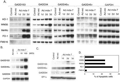
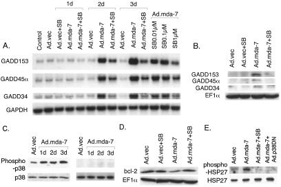
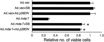
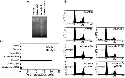
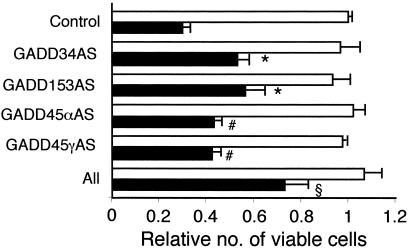
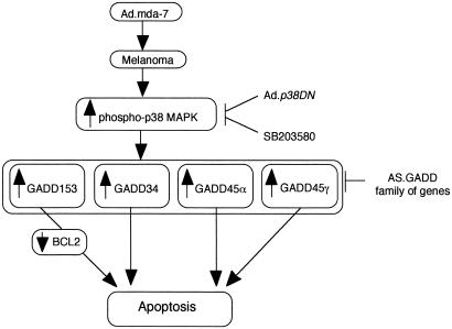
Subtraction hybridization identified melanoma differentiation-associated gene-7 (mda-7) as a gene induced during terminal differentiation in human melanoma cells. On the basis of structure, chromosomal localization and cytokine-like properties, mda-7 is classified as IL-24. Administration of mda-7/IL-24 by means of a replication-incompetent adenovirus (Ad.mda-7) induces apoptosis selectively in diverse human cancer cells without inducing harmful effects in normal fibroblast or epithelial cells. The present studies investigated the mechanism underlying this differential apoptotic effect. Infection of melanoma cells, but not normal immortal melanocytes, with Ad.mda-7 induced a time- and dose-dependent increase in expression, mRNA and protein, of a family of growth arrest and DNA damage (GADD)-inducible genes, which correlated with induction of apoptosis. Among the members of the GADD family of genes, GADD153, GADD45 alpha, and GADD34 displayed marked, and GADD45 gamma showed minimal induction. Treatment of melanoma cells with SB203580, a selective inhibitor of the p38 mitogen-activated protein kinase (MAPK) pathway, effectively inhibited Ad.mda-7-induced apoptosis. Additional support for an involvement of the p38 MAPK pathway in Ad.mda-7-mediated apoptosis was documented by using an adenovirus expressing a dominant negative mutant of p38 MAPK. Infection with Ad.mda-7 increased the phosphorylation of p38 MAPK and heat shock protein 27 in melanoma cells but not in normal immortal melanocytes. In addition, SB203580 effectively inhibited Ad.mda-7-mediated induction of the GADD family of genes in a time- and dose-dependent manner, and it effectively blocked Ad.mda-7-mediated down-regulation of the antiapoptotic protein BCL-2. Inhibition of GADD genes by an antisense approach either alone or in combination also effectively blocked Ad.mda-7-induced apoptosis in melanoma cells. These results support the hypothesis that Ad.mda-7 mediates induction of the GADD family of genes by means of the p38 MAPK pathway, thereby resulting in the selective induction of apoptosis in human melanoma cells.


Figures

Figure 1
Figure 1
Infection with Ad.mda-7 induces the GADD family of genes in melanoma cells but not in normal immortal melanocytes in a time- and dose-dependent manner. (A) Melanoma cells (HO-1, WM35, MeWo, and FO-1) and immortalized human melanocytes (FM516) were infected with either Ad.vec or with Ad.mda-7 at a multiplicity of infection (MOI) of 100 pfu per cell for 3 days. Total RNA was extracted and Northern analysis was performed by using the indicated cDNA probes as described in Materials and Methods. (B) FO-1 cells were infected with either Ad.vec (100 pfu per cell) or Ad.mda-7 (1, 10, and 100 pfu per cell) and Northern blot analysis was performed as indicated. (C) FO-1 cells were infected with either Ad.vec or Ad.mda-7 (100 pfu per cell) for 3 days. Cell lysates were prepared and Western blot analysis was performed by using the indicated antibodies as described in Materials and Methods. (D) Percentage of cells displaying hypodiploidy (Ao), a measure of apoptosis, by fluorescence-activated cell sorter analysis 3 days after infection with a multiplicity of infection of 100 pfu per cell of Ad.mda-7.
Figure 2
Figure 2
Treatment with SB203580 inhibits Ad.mda-7-mediated induction of the GADD family of genes, p38 MAPK phosphorylation, and BCL-2 protein down-regulation. (A) FO-1 cells were infected with either Ad.vec or with Ad.mda-7 (100 pfu per cell) and were treated with either 1 μM SB203580 for 3 days or with different concentrations of SB203580 for 2 days. Total RNA was extracted and Northern blot analysis was performed. (B) FO-1 cells were infected with either Ad.vec or Ad.mda-7 (100 pfu per cell) and treated with 1 μM SB203580 for 3 days. Cell lysates were prepared and Western blot analysis was performed. (C) FO-1 (Left) and FM516 (Right) cells were infected with either Ad.vec or Ad.mda-7 (100 pfu per cell) for 3 days. Cell lysates were prepared and Western blot analysis was performed with anti-phospho-p38 and anti-p38 MAPK antibodies as described in Materials and Methods. (D) FO-1 cells were infected with either Ad.vec or Ad.mda-7 (100 pfu per cell) and were treated with 1 μM SB203580 for 3 days. Cell lysates were prepared and Western blot analysis was performed by using the indicated antibodies. (E) FO-1 cells were infected with either Ad.vec or Ad.mda-7 (100 pfu per cell) and were either treated with 1 μM SB203580 or infected with dominant negative p38 MAPK (Ad.p38DN; 100 pfu per cell) for 3 days. Cell lysates were prepared and Western blot analysis was performed by using the indicated antibodies.
Figure 3
Figure 3
Inhibition of the p38 MAPK pathway protects FO-1 melanoma cells from Ad.mda-7-mediated cell death. FO-1 cells were infected with either Ad.vec or Ad.mda-7 (100 pfu per cell) and treated with 1 μM SB203580 or infected with Ad.p38DN (100 pfu per cell). Cell viability was measured by MTT assay after 4 days. Cell viability of Ad.vec-treated cells was regarded as 1. *, significant differences from Ad.mda-7 (P < 0.0001).
Figure 4
Figure 4
Inhibition of the p38 MAPK pathway protects cells from Ad.mda-7-mediated apoptosis. (A) FO-1 cells were infected with either Ad.vec or Ad.mda-7 (100 pfu per cell) and treated with 1 μM SB203580 for 3 days. DNA was isolated from the cells and fragmentation was analyzed as described in Materials and Methods. (B) FO-1 cells were infected with either Ad.vec or with Ad.mda-7 (100 pfu per cell) and treated with 1 μM SB203580 or infected with Ad.p38DN (100 pfu per cell). Cell cycle was analyzed as described in Materials and Methods. (C) FO-1 cells were infected with either Ad.vec or with Ad.mda-7 (100 pfu per cell) and treated with 1 μM SB203580 or infected with Ad.p38DN (100 pfu per cell). Percentage of apoptotic cells at days 1 and 3 after infection in each group were plotted.
Figure 5
Figure 5
Inhibition of the GADD family of genes protects FO-1 melanoma cells from Ad.mda-7-mediated cell death. FO-1 cells were transfected with the indicated antisense construct either alone or in combination. After 24 h the cells were infected with either Ad.vec (white bars) or Ad.mda-7 (black bars) (100 pfu per cell) for 3 days. Cell viability was measured by MTT assay. Cell viability of Ad.vec-treated cells was regarded as 1. Significant differences from Control, Ad.mda-7 #, P < 0.05; *, P < 0.005; §, P < 0.0001.
Figure 6
Figure 6
A hypothetical model of the involvement of the p38 MAPK pathway and the GADD family members of genes in mediating apoptosis in human melanoma cells by Ad.mda-7.

Similar articles

Cited by

References

    1. Sachs L. Nature (London) 1978;274:535–539. - PubMed
    1. Fisher P B, Grant S. Pharmacol Ther. 1985;27:143–166. - PubMed
    1. Jiang H, Lin J, Fisher P B. Mol Cell Differ. 1994;2:221–239.
    1. Leszczyniecka M, Roberts T, Dent P, Grant S, Fisher P B. Pharmacol Ther. 2001;90:105–156. - PubMed
    1. Waxman S. Differentiation Therapy. Rome: Ares-Serono Symposia Publishers; 1996. pp. 1–531.

Publication types

MeSH terms

Substances