Regulation of squalene synthase, a key enzyme of sterol biosynthesis, in tobacco
- PMID: 12114564
- PMCID: PMC166504
- DOI: 10.1104/pp.001438
Regulation of squalene synthase, a key enzyme of sterol biosynthesis, in tobacco
Abstract
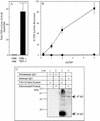
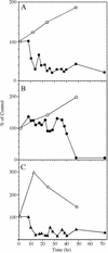
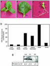
Squalene synthase (SS) represents a putative branch point in the isoprenoid biosynthetic pathway capable of diverting carbon flow specifically to the biosynthesis of sterols and, hence, is considered a potential regulatory point for sterol metabolism. For example, when plant cells grown in suspension culture are challenged with fungal elicitors, suppression of sterol biosynthesis has been correlated with a reduction in SS enzyme activity. The current study sought to correlate changes in SS enzyme activity with changes in the level of the corresponding protein and mRNA. Using an SS-specific antibody, the initial suppression of SS enzyme activity in elicitor-challenged cells was not reflected by changes in the absolute level of the corresponding polypeptide, implicating a post-translational control mechanism for this enzyme activity. In comparison, the absolute level of the SS mRNA did decrease approximately 5-fold in the elicitor-treated cells, which is suggestive of decreased transcription of the SS gene. Study of SS in intact plants was also initiated by measuring the level of SS enzyme activity, the level of the corresponding protein, and the expression of SS gene promoter-reporter gene constructs in transgenic plants. SS enzyme activity, polypeptide level, and gene expression were all localized predominately to the shoot apical meristem, with much lower levels observed in leaves and roots. These later results suggest that sterol biosynthesis is localized to the apical meristems and that apical meristems may be a source of sterols for other plant tissues.
Figures


Similar articles
-
Molecular characterization of tobacco squalene synthase and regulation in response to fungal elicitor.Arch Biochem Biophys. 1998 Jan 15;349(2):205-15. doi: 10.1006/abbi.1997.0463. Arch Biochem Biophys. 1998. PMID: 9448707
-
Molecular cloning, bacterial expression and promoter analysis of squalene synthase from Withania somnifera (L.) Dunal.Gene. 2012 May 10;499(1):25-36. doi: 10.1016/j.gene.2012.03.004. Epub 2012 Mar 9. Gene. 2012. PMID: 22425978
-
Inhibition of squalene synthase and squalene epoxidase in tobacco cells triggers an up-regulation of 3-hydroxy-3-methylglutaryl coenzyme a reductase.Plant Physiol. 2002 Sep;130(1):334-46. doi: 10.1104/pp.004655. Plant Physiol. 2002. PMID: 12226513 Free PMC article.
-
Squalene synthase: structure and regulation.Prog Nucleic Acid Res Mol Biol. 2001;65:157-95. doi: 10.1016/s0079-6603(00)65005-5. Prog Nucleic Acid Res Mol Biol. 2001. PMID: 11008488 Review.
-
Inhibitors of sterol biosynthesis and their applications.Prog Lipid Res. 1993;32(4):357-416. doi: 10.1016/0163-7827(93)90016-p. Prog Lipid Res. 1993. PMID: 8309949 Review. No abstract available.
Cited by
-
The Sterol Trafficking Pathway in Arabidopsis thaliana.Front Plant Sci. 2021 May 26;12:616631. doi: 10.3389/fpls.2021.616631. eCollection 2021. Front Plant Sci. 2021. PMID: 34122463 Free PMC article.
-
Transcriptome analysis reveals metabolic alteration due to consecutive monoculture and abiotic stress stimuli in Rehamannia glutinosa Libosch.Plant Cell Rep. 2017 Jun;36(6):859-875. doi: 10.1007/s00299-017-2115-2. Epub 2017 Mar 8. Plant Cell Rep. 2017. PMID: 28275853
-
Comparative transcriptome analysis of different tissues of Hylomecon japonica provides new insights into the biosynthesis pathway of triterpenoid saponins.Front Bioinform. 2025 Jul 7;5:1625145. doi: 10.3389/fbinf.2025.1625145. eCollection 2025. Front Bioinform. 2025. PMID: 40693151 Free PMC article.
-
Characterization of Squalene synthase gene from Chlorophytum borivilianum (Sant. and Fernand.).Mol Biotechnol. 2013 Jul;54(3):944-53. doi: 10.1007/s12033-012-9645-1. Mol Biotechnol. 2013. PMID: 23338982
-
Cyclodextrins Increase Triterpene Production in Solanum lycopersicum Cell Cultures by Activating Biosynthetic Genes.Plants (Basel). 2022 Oct 20;11(20):2782. doi: 10.3390/plants11202782. Plants (Basel). 2022. PMID: 36297806 Free PMC article.
References
-
- Adler JH, Grebenok RJ. Biosynthesis and distribution of insect-molting hormones in plants: a review. Lipids. 1995;30:257–262. - PubMed
-
- Bhatt PN, Bhatt DP. Regulation of sterol biosynthesis in Solanum species. J Exp Bot. 1984;35:890–896.
-
- Brindle PA, Kuhn PJ, Threlfall DR. Biosynthesis and metabolism of sesquiterpenoid phytoalexins and triterpenoids in potato cell-suspension cultures. Phytochemistry. 1988;27:133–150.
-
- Brown MS, Goldstein JL. The SREBP pathway: regulation of cholesterol metabolism by proteolysis of a membrane-bound transcription factor. Cell. 1997;89:331–340. - PubMed
-
- Caelles C, Ferrer A, Balcells L, Hegardt FG, Boronat A. Isolation and structural characterization of a cDNA-encoding Arabidopsis thaliana 3-hydroxy-3-methylglutaryl coenzyme-A reductase. Plant Mol Biol. 1989;13:627–638. - PubMed
Publication types
MeSH terms
Substances
Associated data
- Actions
LinkOut - more resources
Full Text Sources
Other Literature Sources

