Comparative Study
. 2002 Jul;129(3):1181-93.
doi: 10.1104/pp.011024.

Leaf senescence and starvation-induced chlorosis are accelerated by the disruption of an Arabidopsis autophagy gene

Affiliations
Comparative Study

Leaf senescence and starvation-induced chlorosis are accelerated by the disruption of an Arabidopsis autophagy gene

Hideki Hanaoka et al. Plant Physiol. 2002 Jul.

Abstract

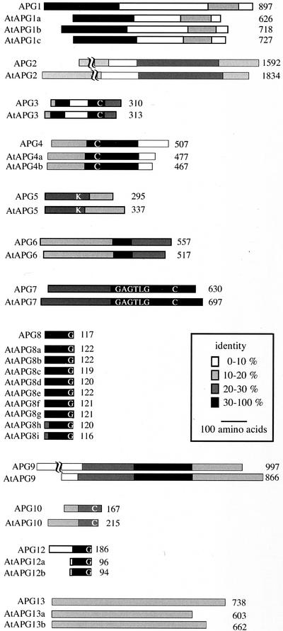
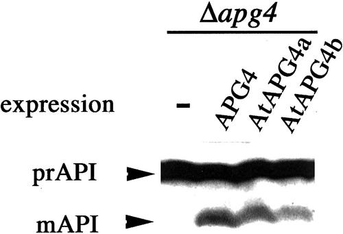
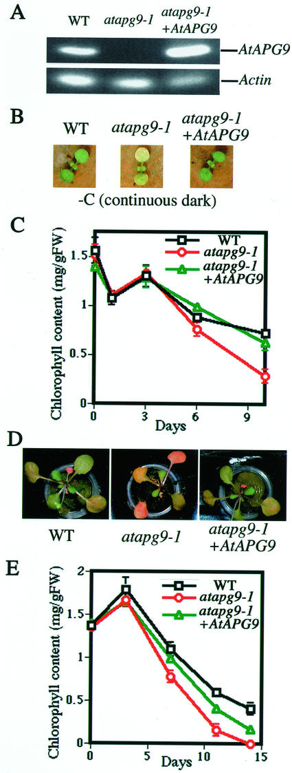
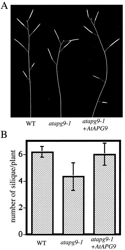
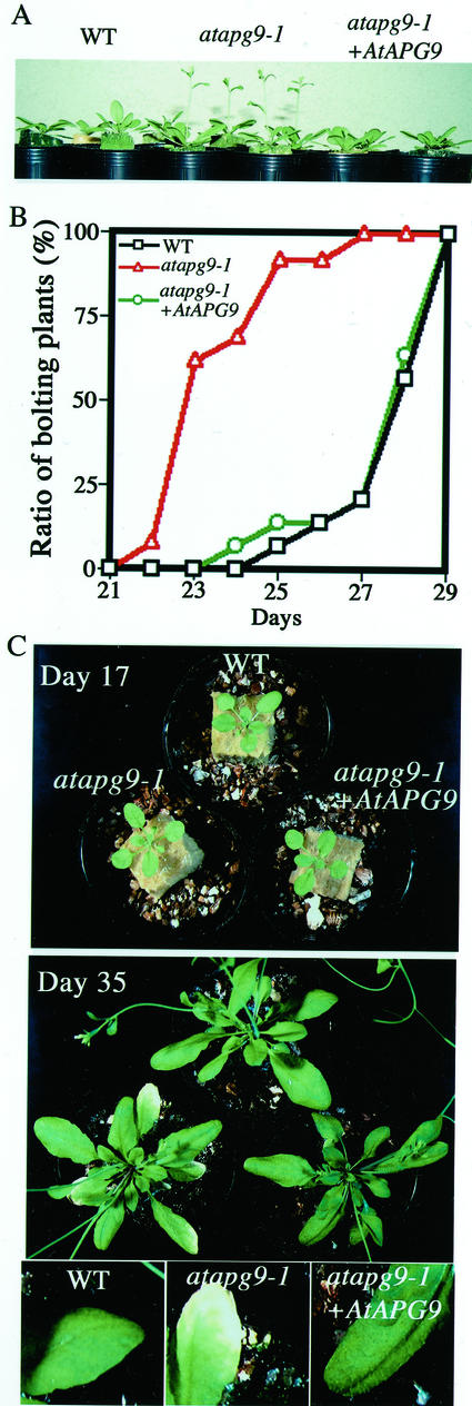
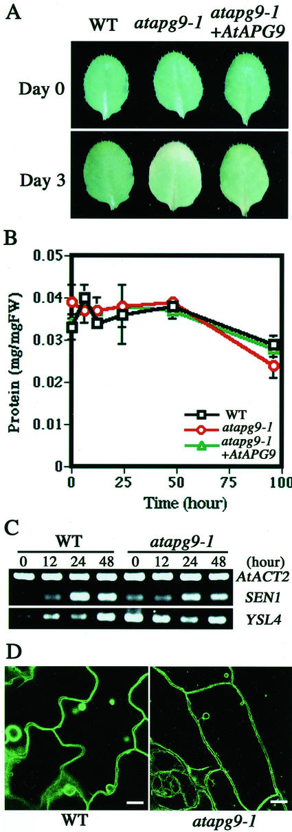
Autophagy is an intracellular process for vacuolar bulk degradation of cytoplasmic components. The molecular machinery responsible for yeast and mammalian autophagy has recently begun to be elucidated at the cellular level, but the role that autophagy plays at the organismal level has yet to be determined. In this study, a genome-wide search revealed significant conservation between yeast and plant autophagy genes. Twenty-five plant genes that are homologous to 12 yeast genes essential for autophagy were discovered. We identified an Arabidopsis mutant carrying a T-DNA insertion within AtAPG9, which is the only ortholog of yeast Apg9 in Arabidopsis (atapg9-1). AtAPG9 is transcribed in every wild-type organ tested but not in the atapg9-1 mutant. Under nitrogen or carbon-starvation conditions, chlorosis was observed earlier in atapg9-1 cotyledons and rosette leaves compared with wild-type plants. Furthermore, atapg9-1 exhibited a reduction in seed set when nitrogen starved. Even under nutrient growth conditions, bolting and natural leaf senescence were accelerated in atapg9-1 plants. Senescence-associated genes SEN1 and YSL4 were up-regulated in atapg9-1 before induction of senescence, unlike in wild type. All of these phenotypes were complemented by the expression of wild-type AtAPG9 in atapg9-1 plants. These results imply that autophagy is required for maintenance of the cellular viability under nutrient-limited conditions and for efficient nutrient use as a whole plant.


Figures

Figure 1
Figure 1
Comparison of yeast Apg proteins and Arabidopsis AtAPG proteins. The shading indicates the degree of identity between each homologous region. Each protein was aligned based on the CLUSTAL V method using DNASTAR. Conserved amino acid residues important for proper Apg function are indicated by letters. The numbers indicate the amino acid length of each protein. Accession numbers for the AtAPG sequences are: AtAPG1a, AAK59554; AtAPG3, AB073170; AtAPG4a, AB073171; AtAPG4b, AB073172; AtAPG5, AI997825; AtAPG6, AAK62668; AtAPG7, AB073173; AtAPG8a, AB073175; AtAPG8b, AB073176; AtAPG8c, AB073177; AtAPG8d, AB073178; AtAPG8e, AB073179; AtAPG8f, AB073180; AtAPG8g, AB073181; AtAPG8h, AB073182; AtAPG8i, AB073183; AtAPG9, AB073174; AtAPG12a, AB073184; and AtAPG12b, AB073185. The rest of the AtAPG protein sequences are predicted based upon genome sequence. Accession numbers for the corresponding BAC clones are: AtAPG1b, AL132960; AtAPG1c, AC007661; AtAPG2, AP000419; AtAPG10, AC009853; AtAPG13a, AL132964; and AtAPG13b, AB026654.
Figure 2
Figure 2
Complementation of yeast apg4 mutants by AtApg4. Protein extracts from yeast Δapg4 mutant cells or those expressing yeast APG4, Arabidopsis AtAPG4a, or AtAPG4b were analyzed by immunoblot using antiserum against API. The positions of precursor and mature API are indicated.
Figure 3
Figure 3
Amino acid alignment of AtAPG9 and its orthologs. Each protein was aligned by the CLUSTAL V method using DNASTAR. Residues that match the consensus are shaded in black. At, Arabidopsis; Sc, yeast; Ce, Caenorhabditis elegans; Dm, fruitfly; and Hs, human (Homo sapiens).
Figure 4
Figure 4
Identification of the mutant atapg9-1. A, Genomic structure of the AtAPG9 gene. Lines indicate introns and boxes indicate exons; white boxes, untranslated regions; and black boxes, translated regions. The T-DNA insertion site in the atapg9-1 allele is indicated by the gray box. B, Southern-blot analysis of the AtAPG9 gene. Arabidopsis genomic DNA from wild-type (WT) and atapg9-1 mutant plants was digested with BamHI (Ba), BglII (Bg), or EcoRV (E), and the blot was hybridized with an AtAPG9 probe. C, The expression of AtAPG9 in various organs. Total RNA was isolated from flowers, leaves, stems, and roots of wild-type plants grown hydroponically for 1 month. RT-PCR was performed using gene-specific primers for AtAPG9 and for actin ACT2 gene. After agarose electrophoresis, the gel was stained with ethidium bromide. D, Immunoblotting of AtAPG9 in plant lysates. Total plant lysates prepared from wild-type plants (WT) and atapg9-1 mutant plants were centrifuged at 100,000g for 1 h, and the pellets were subjected to the immunoblot using anti-AtAPG9. Possible degradation product of AtAPG9 is marked by the asterisk. E, Immunoblotting of AtAPG9 in yeast lysates. Yeast total lysates were prepared from yeast Δapg9 cells or Δapg9 cells expressing AtAPG9 as described in “Materials and Methods.” AtAPG9 (predicted molecular mass = 99 kD) is indicated by the arrow.
Figure 5
Figure 5
AtAPG9 expression in atapg9-1 suppresses carbon or nitrogen starvation-induced chlorosis. A, RT-PCR of AtAPG9 gene. Total RNA was isolated from leaves of wild type, atapg9-1, and atapg9-1 transformed with the AtAPG9 gene. RT-PCR was performed using gene-specific primers for AtAPG9 and for actin ACT2 gene. After the agarose electrophoresis, the gel was stained with ethidium bromide. B, Top view of 15-d-old carbon-starved plants. Plants were photographed after 8 d of carbon starvation. C, Time-course analysis of chlorophyll content. Plants were grown for 7 d with a light cycle of 16 h light/8 h dark, after which they were maintained in the dark. Chlorophyll was extracted from two cotyledons at the day indicated after transfer to continuous dark conditions. D, Top view of 24-d-old nitrogen-starved plants. Plants were grown with nutrient medium containing 7 mm nitrate for 10 d and then transferred to nitrogen-depleted (0 mm nitrate) medium and grown hydroponically for 14 d. E, Time-course analysis of chlorophyll content. Chlorophyll was extracted from the first and second rosette leaves at the day indicated after induction of nitrogen starvation. All measurements were made on at least three individual plants.
Figure 6
Figure 6
AtAPG9 deficiency impairs efficient seed production under nitrogen-starvation conditions. A, Representative 2-month-old plants grown hydroponically under nitrogen-starvation conditions. B, Number of siliques produced per plant grown under nitrogen-starvation conditions for 2 months. All measurements were made on at least four individual plants.
Figure 7
Figure 7
The phenotypes of atapg9-1 under normal condition. A, Side view of 26-d-old plants. Wild-type and atapg9-1 plants were grown at 22°C with a 16-h-light/8-h-dark cycle supplied with standard nutrient solution. B, Time-course analysis of bolting. The number of plants containing at least one primary inflorescence stem longer than 5 mm was counted daily. All measurements were made on at least 13 individual plants. C, Natural leaf senescence. Wild type, atapg9-1, and atapg9-1 transformed with wild-type AtAPG9 gene were grown under standard conditions and photographed on d 17 and 35 after germination. Magnified views were also shown.
Figure 8
Figure 8
The phenotypes during artificially induced senescence. The third or fourth rosette leaves of 3-week-old plants were detached and floated on water at 22°C in the dark. A, Top view of detached leaves. The leaves were photographed at d 0 and after 3 d of incubation. B, Changes in protein content. Protein was extracted from the detached rosette leaves at the time indicated. All measurements were made on at least three individual plants. C, Expression of senescence-inducible genes. Total RNA (10 μg) isolated from wild type or atapg9-1 were isolated at the time indicated. Semiquantitative RT-PCR was performed using gene-specific primers for SEN1, YSL4, and actin ACT2 gene. After the agarose electrophoresis, the gel was stained with ethidium bromide. D, Vacuolar morphology of epidermal cells in detached rosette leaves. Detached rosette leaves of wild-type or mutant plants expressing γ-TIP-GFP were observed after 12 h of incubation in water. Scale bar = 10 μm.

Similar articles

Cited by

References

    1. Aubert S, Gout E, Bligny R, Marty-Mazars D, Barrieu F, Alabouvette J, Marty F, Douce R. Ultrastructural and biochemical characterization of autophagy in higher plant cells subjected to carbon deprivation: control by the supply of mitochondria with respiratory substrates. J Cell Biol. 1996;133:1251–1263. - PMC - PubMed
    1. Brouquisse R, Gaudillere JP, Raymond P. Induction of a carbon-starvation-related proteolysis in whole maize plants submitted to light/dark cycles and to extended darkness. Plant Physiol. 1998;117:1281–1291. - PMC - PubMed
    1. Bushnell TP, Bushnell D, Jagendorf AT. Purified zinc protease of pea chloroplasts, EP1, degrades the large subunit of ribulose-1,5-bisphosphate carboxylase/oxygenase. Plant Physiol. 1993;103:585–591. - PMC - PubMed
    1. Chen MH, Liu LF, Chen YR, Wu HK, Yu SM. Expression of alpha-amylases, carbohydrate metabolism, and autophagy in cultured rice cells is coordinately regulated by sugar nutrient. Plant J. 1994;6:625–636. - PubMed
    1. Chirgwin JJ, Przbyla AE, MacDonald RJ, Rutter WJ. Isolation of biologically active ribonucleic acid from sources enriched in ribonuclease. Biochemistry. 1979;18:5294–5299. - PubMed

Publication types

MeSH terms

Associated data