Comparative Study
. 2002 Jul;129(3):1407-16.
doi: 10.1104/pp.010983.

Differential messenger RNA gradients in the unicellular alga Acetabularia acetabulum. Role of the cytoskeleton

Affiliations
Comparative Study

Differential messenger RNA gradients in the unicellular alga Acetabularia acetabulum. Role of the cytoskeleton

Heiko Vogel et al. Plant Physiol. 2002 Jul.

Abstract

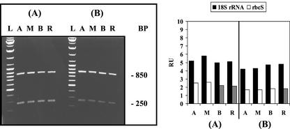
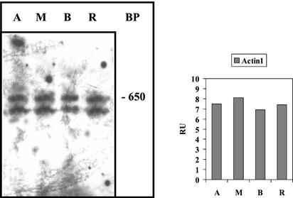
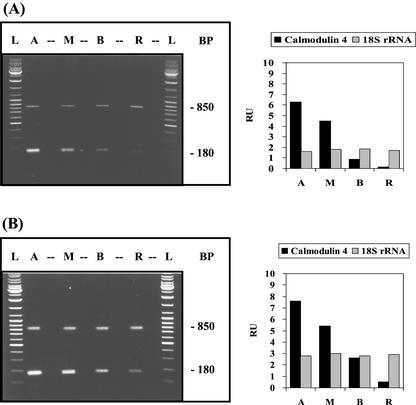
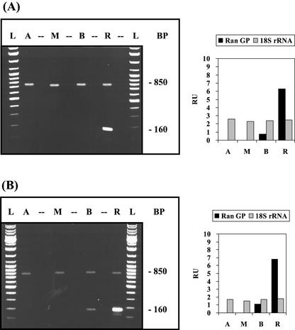
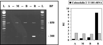
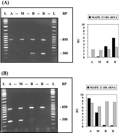
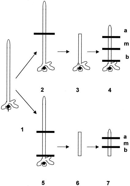
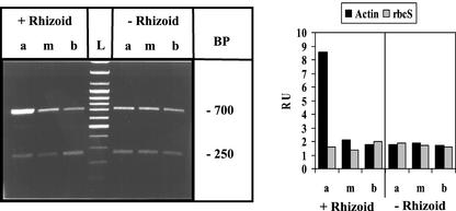
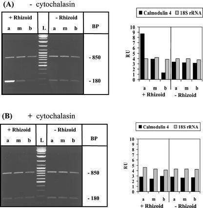
The unicellular green alga Acetabularia acetabulum has proven itself to be a superior model for studies of morphogenesis because of its large size and distinctive polar morphology. The giant cell forms an elongated tube (a stalk of up to 60 mm in length), which at its apical pole makes whorls of hairs, followed by one whorl of gametophores in the shape of a cap. At its basal pole, the cell extends into a rhizoid wherein the single nucleus is positioned. In this study, we have determined the level of specific messenger RNAs in the apical, middle, and basal regions using reverse transcriptase-PCR methodology. Four mRNA classes were distinguished: those that were uniformly distributed (small subunit of Rubisco, actin-1, ADP-glucose, centrin, and alpha- and beta-tubulin), those that expressed apical/basal (calmodulin-4) or basal/apical gradients (calmodulin-2 and a Ran-G protein), and those with development-specific patterns of distribution (mitogen-activated protein kinase, actin-2, and UDP-glucose-epimerase). Restoration of the apical/basal calmodulin-4 mRNA gradient after amputation of the apical region of the cell requires the nucleus and was abolished by cytochalasin D. Accumulation of actin-1 mRNA in the vicinity of the wound set by the amputation needs, likewise, the presence of the nucleus and was also inhibited by cytochalasin. This suggests that actin microfilaments of the cytoskeleton are involved in directed transport and/or anchoring of these mRNAs.


Figures

Figure 1
Figure 1
Distribution of 18S rRNA and rbcS mRNA in different A. acetabulum cell fragments: evaluation by semiquantitative RT-PCR analysis. Young cells of 20 to 30 mm in length without caps (A) and adult cells of 40 to 50 mm in length with nearly full-sized caps (B) were cut into four fragments corresponding to apical stalk fragment (A), middle stalk fragment (M), basal stalk fragment (B), and rhizoidal fragment (R). Equal amounts of total RNA, isolated from these cell fragments, were used as templates in RT-PCR with appropriate specific primers (see “Materials and Methods”). Left panel, Separation of RT-PCR products by agarose gel electrophoresis. The 850-bp piece corresponds to 18S rRNA and the 250-bp piece to rbcS mRNA. (L) indicates a DNA size ladder. Right panel, Densitometric record of the individual signals after blotting and hybridization with fluorescein-labeled A. acetabulum cDNA probes, expressed as relative units (RU).
Figure 2
Figure 2
Cellular distribution of actin isoform-1 mRNA evaluated by RNase protection analysis. Cells of 40 to 50 mm in length with caps were used for cell fragmentation. Left panel, Electrophoretic separation of the products of an RNase protection assay of actin-1 mRNA (see “Materials and Methods”). The 650-bp band and the lower band are protected actin-1 mRNA fragments; they occur because the bacterial polymerases used often produce shorter by-products resulting in generation of two different antisense RNA probes. Right panel, Densitometric record of the combined bands after blotting. Abbreviations are the same as in Figure 1.
Figure 3
Figure 3
Apical/basal gradient of calmodulin-4 mRNA. Developmental stages, experimental conditions, and abbreviations used are similar to Figure 1. The 180-bp RT-PCR product corresponds to calmodulin-4 mRNA and the 850-bp RT-PCR product to 18S rRNA.
Figure 4
Figure 4
Basal/apical distribution of calmodulin-2 mRNA. A. acetabulum cells with nearly full-size caps were used. Left, Electrophoretic separation of RT-PCR products. The 300-bp RT-PCR signal corresponds to calmodulin-2 mRNA and the 850-bp signal to 18S rRNA. Abbreviations and conditions are the same as in Figure 1.
Figure 5
Figure 5
Basal/apical gradient of the mRNA for the small GTP-binding protein Ran. Cell stages, experimental conditions, and abbreviations are similar to Figure 1. The 160-bp RT-PCR product corresponds to the mRNA of the small GTP-binding protein Ran (Ran-GP) and the 850-bp RT-PCR product to 18S rRNA.
Figure 6
Figure 6
Reversal of the gradient of MAP-kinase mRNA after cap formation. Distribution of MAP-kinase mRNA in cells without cap (A) and with nearly fully developed cap (B) was evaluated by RT-PCR analogous to the experiment of Figure 1. The 300-bp signal corresponds to MAP-kinase mRNA, the 850-bp signal to 18S rRNA.
Figure 7
Figure 7
Schematic representation of A. acetabulum cell fragments: culture with and without rhizoid and in absence and presence of cytochalasin D. Cells are not drawn to scale; whorls of hairs are omitted. Amputation is indicated by a horizontal line. 1, Intact cell of 30 to 40 mm in length without cap; 2, amputation of apical cell-region only (5) generation of apical- and rhizoid-amputated fragment. The nucleated (3) and enucleated (6) cell fragments were cultured for 7 d in the absence and presence of cytochalasin D. The cultured cellular fragments were then redissected into apical (a), middle (m), and basal (b) fragments (4, 7) and immediately processed for analysis.
Figure 8
Figure 8
Regeneration after injury induces accumulation of actin-1 mRNA at the apical wound site: the role of the nucleus. Basal cell fragments with or without rhizoid were generated and cultured (in the absence of inhibitor) for 3 d after amputation before processing occurred, as described schematically in Figure 7. Experimental conditions are similar to the one described in Figure 1; abbreviations as in Figure 7. The 700-bp RT-PCR product corresponds to actin-1 mRNA and the 250-bp product to rbcS mRNA.
Figure 9
Figure 9
Restablishment of the apical/basal gradient of calmodulin-4 mRNA after decapitation: the role of actin microfilaments. Basal cell fragments (with or without rhizoid) were generated and cultured in the absence or presence of 10 μg mL−1 of cytochalasin D for 7 d after amputation before processing occurred, as described schematically in Figure 7. Experimental conditions are similar to those described in Figure 1; same abbreviations as in Figure 8. The 180-bp RT-PCR product corresponds to calmodulin 4 mRNA and the 850-bp RT-PCR product to 18S rRNA.

Similar articles

Cited by

References

    1. Bachmann P, Zetsche K. A close temporal and spatial correlation between cell growth, cell wall synthesis and the activity of enzymes of mannan synthesis in Acetabularia mediterranea. Planta. 1979;145:331–337. - PubMed
    1. Bachs O, Agell N, Carafoli E. Calmodulin and calmodulin binding proteins in the nucleus. Cell Calcium. 1994;16:289–296. - PubMed
    1. Bashirullah A, Cooperstock RL, Lipshitz HD. RNA localization in development. Annu Rev Biochem. 1998;67:335–394. - PubMed
    1. Berger S, Kaever MJ. Dasycladales. Stuttgart, Germany: Georg Thieme Verlag; 1992.
    1. Berger S, Wittke W, Traub P. Occurrence of proteinaceous 10 nm filaments throughout the cytoplasm of algae of the order Dasycladales. Exp Cell Res. 1998;240:176–186. - PubMed

Publication types

MeSH terms

LinkOut - more resources