. 2002 Aug;51(2):155-63.
doi: 10.1136/gut.51.2.155.

Defining the roles of perforin, Fas/FasL, and tumour necrosis factor alpha in T cell induced mucosal damage in the mouse intestine

Affiliations

Defining the roles of perforin, Fas/FasL, and tumour necrosis factor alpha in T cell induced mucosal damage in the mouse intestine

M Merger et al. Gut. 2002 Aug.

Abstract

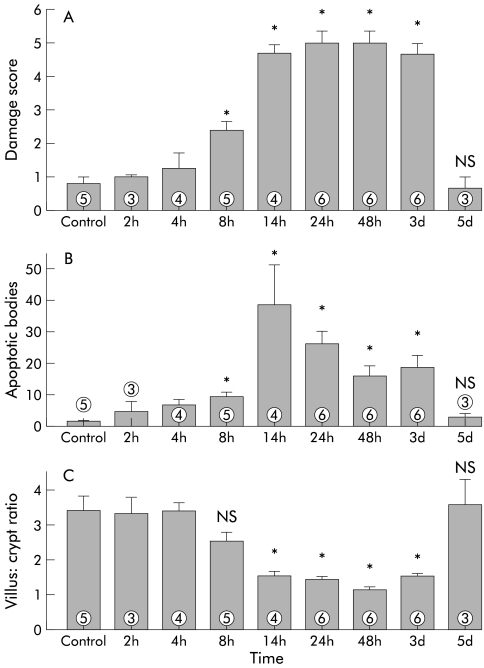
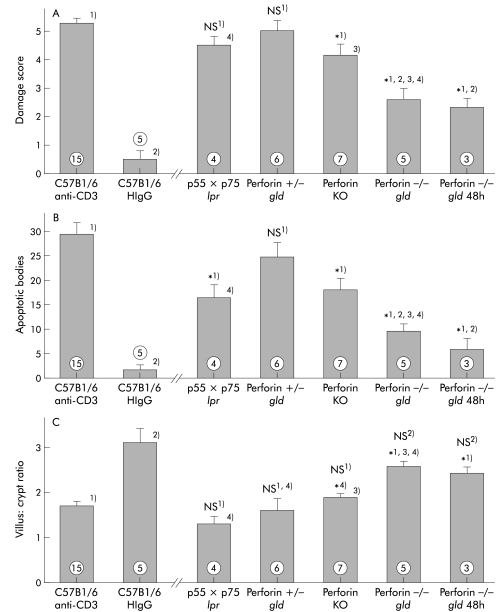
Background and aims: Mucosal flattening and epithelial cell apoptosis are typical features of T cell induced inflammatory diseases of the bowel, such as coeliac disease and graft versus host disease. Mice injected with a T cell activating anti-CD3 antibody develop a severe diarrhoeal illness. We describe the histological features of this enteropathy and define the effector mechanisms involved in T cell induced mucosal injury in this in vivo model.

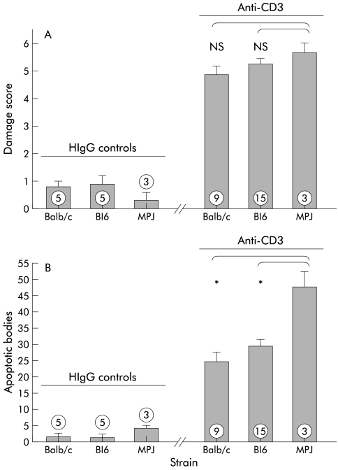
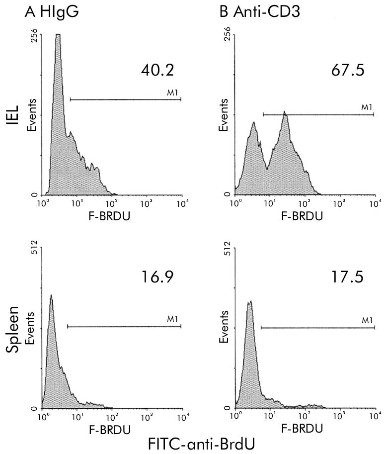
Methods: Wild-type and genetically modified mice were injected with the anti-CD3 antibody 3C11 (50 microg). Changes in the murine intestine were characterised by light microscopy analysis and terminal uridine nick-end labelling (TUNEL) assay. The role of perforin, Fas/Fas ligand (FasL), tumour necrosis factor alpha (TNF-alpha), and interferon gamma (IFN-gamma) in T cell induced mucosal damage was assessed using selected immunodeficient mouse strains.

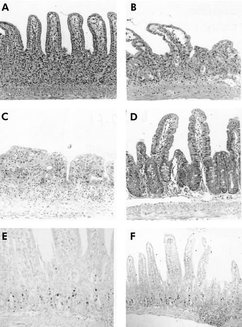
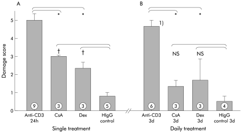
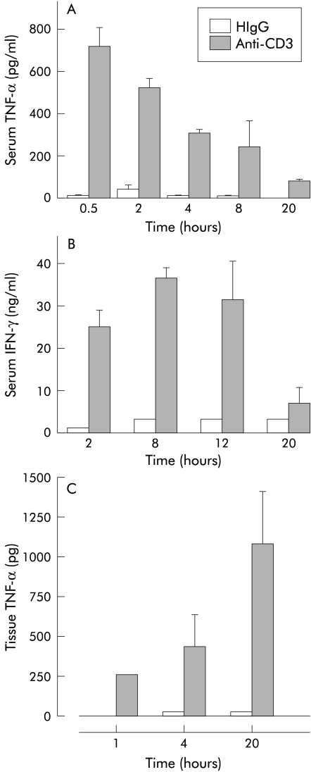
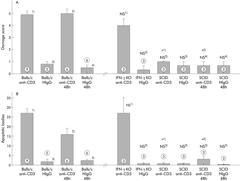
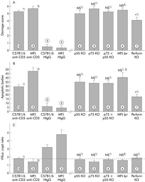
Results: T cell activation caused severe damage, including small intestinal mucosal flattening and apoptosis of crypt epithelial cells. Mucosal damage was unaltered in anti-CD3 treated mice lacking IFN-gamma, Fas, or TNF-alpha receptors. In mice lacking TNF-alpha receptors and Fas (TNF-R1xR2 lpr/lpr strain), enterocyte apoptosis was diminished but there was no significant reduction in tissue damage. Apoptosis and mucosal injury were significantly reduced in perforin knockout mice. Abrogation of both FasL and perforin (perforin KOxgld mice) further significantly reduced tissue damage and apoptotic bodies.

Conclusions: T cell induced mucosal injury is mediated by the combined effect of multiple pathways but predominantly by perforin. The redundancy of the mechanisms of tissue damage will have significant impact on therapeutic strategies aimed at specific and targeted inhibition of inflammatory processes.


Figures

Figure 1
Figure 1
Micrographs of the small intestinal mucosa from Balb/c mice treated with anti-CD3 or hamster IgG control antibody (40×). (A) Mucosa 20 hours after treatment with anti-CD3. The villi are shortened and oedematous; goblet cells and intraepithelial lymphocytes (IEL) are depleted. In the crypt regions, epithelial cells are difficult to distinguish and many apoptotic bodies can be found. Note villus height, short crypts, goblet cells, and IEL. (B) After 40 hours, villi are short and plump. Apoptotic bodies have disintegrated into nuclear fragments. (C) Mucosa 72 hours after anti-CD3 treatment. (D) Five days after anti-CD3 treatment, the mucosa appears normal. Terminal uridine nick-end labelling (TUNEL) assay demonstrates DNA fragmentation as a hallmark of apoptosis of epithelial cells 12 hours after anti-CD3 injection ((E) 40× and (F) 10× magnification).
Figure 2
Figure 2
Time course of small intestinal mucosal damage in Balb/c mice treated with 50 μg of anti-CD3 antibody 145-2C11. (A) A significant increase (p<0.05) in damage score was observed beginning eight hours post injection (data are mean (SEM)). Tissue damage was highly significant (p<0.005) from 24 hours to three days post injection. Tissue damage had completely resolved by five days post injection. The damage score of mice treated with hamster IgG control antibody was below 1 for all time points (data not shown; n=3–6). (B) The number of apoptotic bodies increased significantly after anti-CD3 treatment (p<0.005 at 24 hours to three days post injection). (C) The ratio of villus height to crypt length was significantly reduced by 14 hours post injection (p<0.005 at 24 hours to three days post injection). *p<0.05 compared with controls; NS, no significant difference compared with controls (numbers indicate the number of animals treated).
Figure 3
Figure 3
Damage score of wild-type background strains 24 hours after treatment with either hamster IgG (HIgG) control antibody (left) or anti-CD3 antibody (right). The histological damage that developed in the small intestine 24 hours after treatment with anti-CD3 antibody of C57Bl/6 mice and MPJ mice was similar to that observed in Balb/c mice (A). The number of apoptotic bodies was significantly higher in MPJ mice compared with both Balb/c or C57Bl/6 mice (B). *p<0.05 compared with MPJ mice treated with anti-CD3 antibody (numbers indicate the number of animals treated).
Figure 4
Figure 4
Activation of intraepithelial lymphocytes (IEL) in vivo is shown by demonstration of increased DNA synthesis (incorporation of uridine analogue bromodeoxyuridine (BrdU)). Single colour histograms are shown depicting the proportion of IEL that stained positive with FITC anti-BrdU antibody. (A) IEL isolated from mice treated with hamster IgG (HIgG) control antibody. (B) IEL isolated from mice 24 hours after treatment with anti-CD3 antibody.
Figure 5
Figure 5
Immunosuppressive treatment significantly reduced tissue damage induced by T cell activation. Treatment with a single dose of either cyclosporin A (CsA) or dexamethasone (Dex) significantly reduced the damage score observed 24 hours post injection (A). The damage score in mice treated with daily doses of either CsA or Dex was not different from control mice treated with only hamster IgG (HIgG) control antibody (B). *p<0.05 compared with control mice treated with only anti-CD3 antibody; †p<0.05 compared with control mice treated with only HIgG control antibody; NS, no significant difference compared with mice treated with HIgG control antibody. Damage score was assessed three days post injection (numbers indicate the number of animals treated).
Figure 6
Figure 6
Serum and small intestinal tissue cytokine levels in Balb/c mice injected with anti-CD3 antibody or hamster IgG (HIgG) control antibody. (A) Serum tumour necrosis factor α (TNF-α); (B) serum interferon γ (IFN-γ); (C) mucosal tissue TNF-α levels (n=3).
Figure 7
Figure 7
Histological damage was not reduced in the absence of interferon γ (IFN-γ) but was completely abolished in the absence of T cells. Damage score (A) and number of apoptotic bodies (B) of immunodeficient mice 24 hours and 48 hours after treatment with anti-CD3 control antibody. *p<0.05 compared with wild-type Balb/c mice, 24 hours (1) or 48 hours (2) after treatment with anti-CD3 antibody; NS, no significant difference compared with Balb/c mice, 24 hours (3) or 48 hours (4) after treatment with hamster HIgG (HIgG) control antibody (numbers indicate the number of animals treated).
Figure 8
Figure 8
T cell mediated histological damage and development of apoptotic bodies was partially reduced in the absence of perforin but not in the absence of Fas or tumour necrosis factor α (TNF-α) receptors. The score for small intestinal tissue damage (A), number of apoptotic bodies per five villus crypt units (B), and villus:crypt ratio (C) in mice 24 hours after treatment with anti-CD3 antibody are shown. No significant differences between tissue damage in anti-CD3 treated mice of the background strain and immunodeficient mice (MPJ for Fas deficient MRL lpr mice, C57Bl6 for all other mice) was detectable. Tissue damage was significantly reduced in anti-CD3 treated perforin deficient mice. *p<0.05 compared with C57Bl6 control mice treated with anti-CD3 antibody (1); NS, no significant difference compared with control C57Bl6 mice treated with anti-CD3 antibody (1) or control MPJ mice treated with anti-CD3 antibody (2) (numbers indicate the number of animals treated).
Figure 9
Figure 9
Tumour necrosis factor α (TNF-α), Fas ligand (FasL), and perforin contribute to tissue damage. Tissue damage in anti-CD3 treated mice lacking Fas and both TNF-α receptors (TNF-R1×R2 lpr/lpr) was not statistically diminished (p=0.098). However, the number of apoptotic bodies was significantly reduced (p=0.012). Small intestinal damage was dramatically diminished in mice lacking both perforin and FasL (perforin−/− gld mice, p=0.001). No further increase in damage was seen at 48 hours post injection. FasL deficient mice remained susceptible to tissue damage (perforin+/− gld strain). The score for small intestinal tissue damage (A), number of apoptotic bodies per five villus crypt units (B), and villus:crypt ratio (C) in mice 24 hours after treatment with anti-CD3 antibody are shown. All mice to the right of the break in the X axis were treated with anti-CD3. *1, 2, 3, 4), p<0.05 compared with groups labelled with corresponding numbers; NS no significant difference compared with control C57Bl6 mice treated with anti-CD3 antibody (1) or control C57BL6 mice treated with HIgG antibody (2) (numbers indicate the number of animals treated).

References

    1. MacDonald TT, Spencer J. Evidence that activated mucosal T cells play a role in the pathogenesis of enteropathy in human small intestine. J Exp Med 1988;167:1341–9. - PMC - PubMed
    1. Moss SF, Attia L, Scholes JV, et al. Increased small intestinal apoptosis in coeliac disease. Gut 1996;39:811–17. - PMC - PubMed
    1. Strater J, Wellisch I, Riedl S, et al. CD95 (APO-1/Fas)-mediated apoptosis in colon epithelial cells: a possible role in ulcerative colitis. Gastroenterology 1997;113:16. - PubMed
    1. Yang X, Stennicke HR, Wang B, et al. Granzyme B mimics apical caspases. Description of a unified pathway for trans-activation of executioner caspase-3 and -7. J Biol Chem 1998;273:34278–83. - PubMed
    1. Thiele DL, Eigenbrodt ML, Bryde SE, et al. Intestinal graft-versus-host disease is initiated by donor T cells distinct from classic cytotoxic T lymphocytes. J Clin Invest 1989;84:1947–56. - PMC - PubMed

Publication types

MeSH terms