. 2002 Aug;70(8):4002-8.
doi: 10.1128/IAI.70.8.4002-4008.2002.

Up-regulation of Fas (CD95) and induction of apoptosis in intestinal epithelial cells by nematode-derived molecules

Affiliations

Up-regulation of Fas (CD95) and induction of apoptosis in intestinal epithelial cells by nematode-derived molecules

Akio Kuroda et al. Infect Immun. 2002 Aug.

Abstract

Infection by the intestinal nematode Nippostrongylus brasiliensis induces acceleration of apoptosis in the small intestinal villus epithelial cells in vivo. In the present study, we examined whether worm extract or excretory-secretory product induces apoptosis in the rat intestinal epithelial cell line IEC-6 in vitro. In the presence of worm extract or excretory-secretory product (> or =6 microg/ml), IEC-6 cell growth was significantly suppressed, and there was a concomitant increase in the number of detached cells in culture dishes. Detached cells showed nuclear fragmentation, activation of caspase-3, and specific cleavage of poly(ADP-ribose) polymerase, suggesting that apoptosis was induced in these cells. Semiquantitative reverse transcription-PCR showed that expression of Fas (CD95) mRNA was up-regulated as early as 6 h after addition of excretory-secretory product, while Fas ligand expression and p53 expression were not up-regulated. Fluorescence-activated cell sorter analyses revealed a significant increase in Fas expression and a slight increase in FasL expression in IEC-6 cells cultured in the presence of excretory-secretory product, while control IEC-6 cells expressed neither Fas or FasL. These results indicated that N. brasiliensis worms produce and secrete biologically active molecules that trigger apoptosis in intestinal epithelial cells together with up-regulation of Fas expression, although the mechanism of induction of apoptosis remains to be elucidated.


Figures
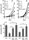
FIG. 1.
FIG. 1.
Effects of WE and ES on the growth of IEC-6 cells. (a and b) IEC-6 cells (0.5 × 10³ cells/well) were cultured in the presence of ES (a) or WE (b) at different doses in 96-well culture dishes, and adherent cell numbers were determined after 1 to 4 days. (c) IEC-6 cells (0.5 × 10³ cells/well) were cultured with 50 μg of untreated WE or ES per ml, 50 μg of heated WE or ES per ml, or 50 μg of proteinase K (Pr-K)-treated WE or ES per ml, and cell numbers were determined after 72 h. The data are means ± standard deviations based on quadruplicate cultures. The results are the results of one of three independent experiments. An asterisk indicates that the P value is <0.001. CNT, control.
FIG. 2.
FIG. 2.
Morphological alterations of IEC-6 cells cultured without ES (A) or with ES (50 μg/ml) (B) for 72 h. In the presence of ES, some cells exhibited shrinkage and long filamentous cytoplasmic projections. Giemsa staining. Magnification, ×100.
FIG. 3.
FIG. 3.
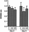
Effects of ES and WE on BrdU incorporation in IEC-6 cells. IEC-6 cells were cultured in the presence or absence of 50 μg of ES per ml or 50 μg of WE per ml for 2 or 3 days and pulsed with BrdU (10 μM) for 2 h before cell harvest. Aliquots (1 × 10⁴ cells) were replated in 96-well plates and fixed, and BrdU levels were determined by an enzyme-linked immunosorbent assay. The data are means + standard deviations based on four wells. The results are the results of one of three independent experiments. C, control; OD450 nm, optical density at 450 nm.
FIG. 4.
FIG. 4.
Short-term effects of WE and ES on IEC-6 cells. (a) Semiconfluent IEC-6 cells were incubated with WE or ES at two doses (10 and 50 μg/ml) for 16 h. Numbers of adherent and detached cells were counted separately as described in Materials and Methods. The percentage of detached cells was calculated as follows: 100 × (number of detached cells)/(number of adherent cells + number of detached cells). The data are averages and standard errors for quadruplicate cultures. Asterisks indicate that values are significantly different from the control culture value (one asterisk, P < 0.05; two asterisks, P < 0.01). (b) Typical apoptotic nuclei in detached cells stained with Hoechst 33258. Magnification, ×100.
FIG. 5.
FIG. 5.
Immunoblot analyses of PARP and caspase-3 in adherent and detached cells. IEC-6 cells were cultured without (−) or with (+) 50 μg of WE per ml for 24 h. Cell lysates of adherent and detached cells were prepared, and the same amounts of protein were loaded and electrophoresed on a 4 to 20% polyacrylamide gradient gel and then transferred onto a nitrocellulose membrane. Immunodetection was carried out with antibodies against PARP and caspase-3.
FIG. 6.
FIG. 6.
Effects of WE or ES on Fas, FasL, and p53 expression in IEC-6 cells. Total RNA isolated from IEC-6 cells was subjected to semiquantitative RT-PCR analyses. (a) To determine appropriate numbers of PCR cycles, the optical densities of the PCR product were measured after different numbers of cycles. Act, β-actin. (b) IEC-6 cells were cultured with or without WE or ES (50 μg/ml) for 48 h, and Fas expression, FasL expression, and p53 expression were analyzed. β-Actin was used as an internal control (CNT). (c) IEC-6 cells were cultured under the same conditions as described above for panel b. The densities of each PCR product were determined with an image analyzer and standardized by using the levels of β-actin. The data are the means + standard errors based on four independent experiments and show expression levels relative to those in control cultures. An asterisk indicates that the level was significantly different from the control level (P < 0.01). (d) IEC-6 cells were cultured with different doses of ES for 48 h, and Fas expression was determined by RT-PCR. (e) IEC-6 cells were cultured with 50 μg of ES per ml, and Fas expression was determined by RT-PCR after different incubation times.
FIG. 7.
FIG. 7.
Effects of heat and proteinase K digestion on ES-induced up-regulation of Fas expression. IEC-6 cells were cultured for 48 h without ES (C1), with heat-inactivated proteinase K (C2), with 50 μg of nontreated ES per ml, with ES after heat treatment, or with ES after proteinase K treatment, and Fas expression was determined by RT-PCR. The data are means + standard errors based on four experiments and show expression levels relative to those of IEC-6 cells cultured for 48 h without ES. Asterisks indicate that the levels are significantly different from control levels (one asterisk, P < 0.05; two asterisks, P < 0.01).
FIG. 8.
FIG. 8.
FACS analyses of Fas and FasL expression on IEC-6 cells. IEC-6 cells were cultured without ES (fine lines) or with 50 μg of ES per ml (thick lines) for 48 h, and harvested cells were incubated with anti-Fas or FasL antibody and then with FITC-conjugated goat anti-rabbit IgG. Shaded area, IEC-6 cells cultured with 50 μg of ES per ml, incubated with normal rabbit serum and then with FITC-conjugated goat anti-rabbit IgG, and subjected to FACS analyses.
FIG. 9.
FIG. 9.
Effects of anti-FasL antibodies on WE-induced cell growth inhibition. IEC-6 cells were cultured with WE (50 μg/ml) in the presence of IgG antibodies against the amino terminus of FasL (N), the carboxy terminus of FasL (C), or normal rabbit IgG (CNT) at different doses, and adherent cell numbers were determined after 72 h. The percentage of inhibition was determined as follows: [1 − (cell numbers in cultures with WE/cell numbers in cultures without WE)] × 100. The data are means + standard deviations based on quadruplicate cultures.
FIG. 10.
FIG. 10.
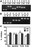
Effects of WE or ES on adherent molecules in IEC-6 cells. (a) IEC-6 cells were cultured with or without WE (50 μg/ml) for 24 h, and β-actin expression (lanes 1 and 2), fibronectin expression (lanes 3 and 4), α2 laminin expression (lanes 5 and 6), β2 laminin expression (lanes 7 and 8), γ1 laminin expression (lanes 9 and 10), and γ2 laminin expression (lanes 11 and 12) were examined by RT-PCR (28 PCR cycles for all experiments except the β-actin experiment, in which 20 cycles were used). C, control; W, WE. (b) IEC-6 cells were cultured with or without WE or ES (50 μg/ml) for 72 h, and β-actin expression (lanes 1 to 3), E-cadherin expression (lanes 4 to 6), and β-catenin expression (lanes 7 to 9) were examined by RT-PCR (28 PCR cycles for all experiments except the β-actin experiment, in which 20 cycles were used). C, control; W, WE; E, ES. (c) IEC-6 cells were cultured with WE or ES (50 μg/ml) in the presence or absence of 100 μg aprotinin per ml, 10 μM TLCK, or 10 μM E-64, and adherent cell numbers were determined after 72 h. The percentage of inhibition was determined as follows: [1 − (cell numbers in cultures with WE or ES/cell numbers in cultures without WE or ES)] × 100. The data are means + standard deviations based on quadruplicate cultures. Non, no aprotinin, TLCK, or E-64 added.

Similar articles

Cited by

References

    1. Appella, E., and C. W. Anderson. 2000. Signaling to p53: breaking the posttranslational modification code. Pathol. Biol. 48:227-245. - PubMed
    1. Batra, S., S. P. Singh, S. Gupta, J. C. Katiyar, and V. M. Srivastava. 1990. Reactive oxygen intermediates metabolizing enzymes in Ancylostoma ceylanicum and Nippostrongylus brasiliensis. Free Radical Biol. Med. 8:271-274. - PubMed
    1. Boudreau, N., C. J. Sympson, Z. Werb, and M. J. Bissell. 1995. Suppression of ICE and apoptosis in mammary epithelial cells by extracellular matrix. Science 267:891-893. - PMC - PubMed
    1. Burt, J. S., and B. M. Ogilvie. 1975. In vitro maintenance of nematode parasites assayed by acetylcholinesterase and allergen secretion. Exp. Parasitol. 38:75-82. - PubMed
    1. Chen, X. M., G. J. Gores, C. V. Paya, and N. F. LaRusso. 1999. Cryptosporidium parvum induces apoptosis in biliary epithelia by a Fas/Fas ligand-dependent mechanism. Am. J. Physiol. 277:g599-608. - PubMed

Publication types