Modification of the structure and activity of lipid A in Yersinia pestis lipopolysaccharide by growth temperature
- PMID: 12117916
- PMCID: PMC128165
- DOI: 10.1128/IAI.70.8.4092-4098.2002
Modification of the structure and activity of lipid A in Yersinia pestis lipopolysaccharide by growth temperature
Abstract
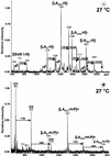
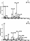
Yersinia pestis strain Yreka was grown at 27 or 37 degrees C, and the lipid A structures (lipid A-27 degrees C and lipid A-37 degrees C) of the respective lipopolysaccharides (LPS) were investigated by matrix-assisted laser desorption ionization-time-of-flight (MALDI-TOF) mass spectrometry. Lipid A-27 degrees C consisted of a mixture of tri-acyl, tetra-acyl, penta-acyl, and hexa-acyl lipid A's, of which tetra-acyl lipid A was most abundant. Lipid A-37 degrees C consisted predominantly of tri- and tetra-acylated molecules, with only small amounts of penta-acyl lipid A; no hexa-acyl lipid A was detected. Furthermore, the amount of 4-amino-arabinose was substantially higher in lipid A-27 degrees C than in lipid A-37 degrees C. By use of mouse and human macrophage cell lines, the biological activities of the LPS and lipid A preparations were measured via their abilities to induce production of tumor necrosis factor alpha (TNF-alpha). In both cell lines the LPS and the lipid A from bacteria grown at 27 degrees C were stronger inducers of TNF-alpha than those from bacteria grown at 37 degrees C. However, the difference in activity was more prominent in human macrophage cells. These results suggest that in order to reduce the activation of human macrophages, it may be more advantageous for Y. pestis to produce less-acylated lipid A at 37 degrees C.
Figures


Similar articles
-
Variation in lipid A structure in the pathogenic yersiniae.Mol Microbiol. 2004 Jun;52(5):1363-73. doi: 10.1111/j.1365-2958.2004.04059.x. Mol Microbiol. 2004. PMID: 15165239
-
Humanized TLR4/MD-2 mice reveal LPS recognition differentially impacts susceptibility to Yersinia pestis and Salmonella enterica.PLoS Pathog. 2012;8(10):e1002963. doi: 10.1371/journal.ppat.1002963. Epub 2012 Oct 11. PLoS Pathog. 2012. PMID: 23071439 Free PMC article.
-
Characterization of the lipopolysaccharide of Yersinia pestis.Microb Pathog. 2001 Feb;30(2):49-57. doi: 10.1006/mpat.2000.0411. Microb Pathog. 2001. PMID: 11162185
-
Structural features and structural variability of the lipopolysaccharide of Yersinia pestis, the cause of plague.J Endotoxin Res. 2006;12(1):3-9. doi: 10.1179/096805105X67283. J Endotoxin Res. 2006. PMID: 16420739 Review.
-
Lipopolysaccharides of bacterial pathogens from the genus Yersinia: a mini-review.Biochimie. 2003 Jan-Feb;85(1-2):145-52. doi: 10.1016/s0300-9084(03)00005-1. Biochimie. 2003. PMID: 12765784 Review.
Cited by
-
Pathogenicity and virulence of Yersinia.Virulence. 2024 Dec;15(1):2316439. doi: 10.1080/21505594.2024.2316439. Epub 2024 Feb 22. Virulence. 2024. PMID: 38389313 Free PMC article. Review.
-
Recent findings regarding maintenance of enzootic variants of Yersinia pestis in sylvatic reservoirs and their significance in the evolution of epidemic plague.Vector Borne Zoonotic Dis. 2010 Jan-Feb;10(1):85-92. doi: 10.1089/vbz.2009.0043. Vector Borne Zoonotic Dis. 2010. PMID: 20158336 Free PMC article.
-
Shield as signal: lipopolysaccharides and the evolution of immunity to gram-negative bacteria.PLoS Pathog. 2006 Jun;2(6):e67. doi: 10.1371/journal.ppat.0020067. PLoS Pathog. 2006. PMID: 16846256 Free PMC article. Review. No abstract available.
-
Discordance in the effects of Yersinia pestis on the dendritic cell functions manifested by induction of maturation and paralysis of migration.Infect Immun. 2006 Nov;74(11):6365-76. doi: 10.1128/IAI.00974-06. Epub 2006 Aug 21. Infect Immun. 2006. PMID: 16923789 Free PMC article.
-
Evaluation of imipenem for prophylaxis and therapy of Yersinia pestis delivered by aerosol in a mouse model of pneumonic plague.Antimicrob Agents Chemother. 2014 Jun;58(6):3276-84. doi: 10.1128/AAC.02420-14. Epub 2014 Mar 31. Antimicrob Agents Chemother. 2014. PMID: 24687492 Free PMC article.
References
-
- Aussel, L., H. Thérisod, D. Karibian, M. B. Perry, M. Bruneteau, and M. Caroff. 2000. Novel variation of lipid A structures in strains of different Yersinia species. FEBS Lett. 465:87-92. - PubMed
-
- Bordet, C., M. Bruneteau, and G. Michel. 1977. Lipopolysaccharides d'une souche à virulence atténuée de Yersinia pestis. Eur. J. Biochem. 79:443-449. - PubMed
MeSH terms
Substances
LinkOut - more resources
Full Text Sources
Other Literature Sources

