. 2002 Aug;70(8):4148-57.
doi: 10.1128/IAI.70.8.4148-4157.2002.

Mycobacterium bovis BCG-infected mice are more susceptible to staphylococcal enterotoxin B-mediated toxic shock than uninfected mice despite reduced in vitro splenocyte responses to superantigens

Affiliations

Mycobacterium bovis BCG-infected mice are more susceptible to staphylococcal enterotoxin B-mediated toxic shock than uninfected mice despite reduced in vitro splenocyte responses to superantigens

João A Pedras-Vasconcelos et al. Infect Immun. 2002 Aug.

Abstract

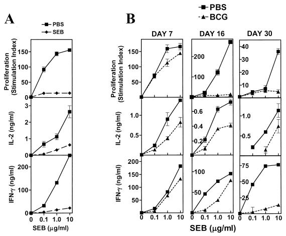
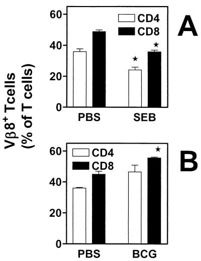
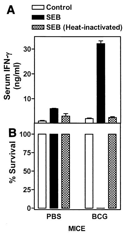
Type 1 T-cell responses against intracellular pathogens play a crucial role in mediating protection. We examined whether the induction of a strong type 1 T-cell response during a chronic bacterial infection influences responses to superantigens capable of inducing acute shock. Intravenous infection of mice with Mycobacterium bovis BCG appeared to induce a progressive anergy towards staphylococcal enterotoxin B (SEB) and towards antigen preparation of BCG (BCG-Ag) itself, based on diminished gamma interferon (IFN-gamma) production by SEB- and BCG-Ag-stimulated splenocytes from infected mice. In contrast to these in vitro results, injection of SEB into BCG-infected mice led to a dramatic increase in the serum IFN-gamma levels and the death of infected but not of control mice. In vitro hyporesponsiveness towards SEB and BCG-Ag occurred only with unfractionated splenocyte cultures, as purified T cells from infected mice produced higher levels of IFN-gamma. Hyporesponsiveness towards SEB and BCG-Ag in unfractionated splenocyte cultures was not due to suppressive antigen-presenting cells (APCs), as APCs from infected mice stimulated higher levels of IFN-gamma from purified T cells. The diminished IFN-gamma levels observed with bulk splenocytes appear to be due to changes in the T-cell-to-APC ratio that result in a decreased proportion of T cells, coupled to reduced proliferative responses and an increased susceptibility of effector T cells to activation-induced cell death in vitro. Our results indicate that the reported phenomena of T-cell anergy during mycobacterial infection may be an in vitro consequence of the development of a strong type 1 response in vivo.


Figures

FIG. 1.
FIG. 1.
BCG infection induces splenic hyporesponsiveness to SEB and mimics in vivo exposure to the superantigen. Female BALB/c mice were either injected with SEB (A), infected with BCG (Pasteur) i.v. (B), or injected with PBS (details in Materials and Methods). On day 7 post-SEB injection or day 7, 16, or 30 post-BCG infection, two or three mice were killed per group and single-cell suspensions were prepared from their spleens. Unfractionated splenocytes were stimulated with various concentrations of SEB (0, 0.1, 1.0, and 10 μg/ml) at 5 × 10⁵ cells/well. Culture conditions, as well as cytokine levels and proliferation (stimulation index) determination, were done as described in Materials and Methods. Mean ± standard deviation of triplicate wells is shown.
FIG. 2.
FIG. 2.
Infection with BCG results in accumulation and not deletion of superantigen-responsive CD4+ or CD8+ T cells. Unfractionated splenocytes (5 × 10⁵ cells/tube) from SEB-exposed mice (7 days postinjection) (A) and PBS-injected controls or day-16-infected mice and PBS-injected controls (B) were stained with FITC-labeled anti-mouse Vβ8 TCR (A and B) and either PE-labeled anti-mouse CD4 or anti-mouse CD8. The percentages of Vβ8+ CD4+ T cells (white bars) or Vβ8+ CD8+ T cells (black bars) in the spleen were determined by flow cytometry. Means ± standard errors of the means of three experiments are shown. ∗, P < 0.05.
FIG. 3.
FIG. 3.
BCG-infected mice have increased levels of IFN-γ in their serum after SEB injection and are more susceptible to SEB-mediated toxic shock. PBS controls and BCG-infected mice were injected with heat-inactivated or normal SEB (100 μg/mouse, three mice/group) i.v. 30 days postinfection. Injected mice, along with uninjected controls, were bled 8 h postinjection, and serum IFN-γ levels were determined by ELISA (A). Animals were monitored but were allowed to progress to terminal shock (B). Experiments were performed twice.
FIG. 4.
FIG. 4.
Purified CD4+ T cells from infected mice are potent IFN-γ producers. (A) Purified CD4+ (>90% pure) from spleens of mice infected at week 2 or of PBS-injected controls were cocultured at 105 cells/well with irradiated bone marrow-derived dendritic cells (5 × 10⁴ cells/well) with various concentrations of SEB. IL-2, IFN-γ, and proliferation (stimulation indexes) were determined as described in Materials and Methods. Mean ± standard deviation of triplicate wells is shown. (B) Unfractionated splenocytes from mice infected for 2 weeks with BCG HD (106 CFU/mouse) or LD (104 CFU/mouse) or from PBS-injected controls were stimulated with various concentrations of SEB or BCG-Ag at 5 × 10⁵ cells/well. Purified CD4+ T cells from each type of spleen were cocultured at 105 cells/well with irradiated bone marrow-derived dendritic cells (5 × 10⁴ cells/well) in the presence of SEB (10 μg/ml) or BCG-Ag (10 μg/ml). IFN-γ levels were determined as described in Materials and Methods. Mean ± standard deviation of triplicate wells is shown.
FIG. 5.
FIG. 5.
BCG infectious dose correlates with increased expression of the T-cell activation markers CD44, IL-2Rα, and IL-2Rβ. Unfractionated splenocytes (5 × 10⁵ cells/tube) from each type of spleen were stained with FITC-labeled anti-mouse CD44, anti-mouse CD25/IL-2Rα, or anti-mouse CD122/IL-2Rβ and PE-labeled anti-mouse CD4 or anti-mouse CD8α. The percentages of CD4+ CD44hi, IL-2Rαhi, or IL-2Rβhi CD4+ (white bars) or CD8+ cells (black bars) in the spleen were determined by flow cytometry. Means ± standard errors of the means of three experiments are shown. ∗, P < 0.05; ∗∗, P < 0.01. Purified CD4+ T cells from the spleens of BCG HD-infected mice (day 16) were stained with FITC anti-mouse CD44 and were sorted in an EPICS Elite ESP (Beckman Coulter) into CD44low (naïve) and CD44hi (memory/effector) populations. Sorted cells were cocultured at 105 cells/well either with bone marrow-derived dendritic cells (5 × 10⁴ cells/well) in the presence or absence of SEB (10 μg/ml) or with M12 B-lymphoma cells (5 × 10⁴ cells/well) in the presence or absence of BCG-Ag (10 μg/ml). IFN-γ levels were determined as described in Materials and Methods. Mean ± standard deviation of triplicate wells is shown.
FIG. 6.
FIG. 6.
BCG infection induces potent APCs that stimulate rather than inhibit IFN-γ production to SEB and BCG-Ag by purified CD4+ T cells. Sixteen days after injection of mice with PBS or BCG LD or BCG HD, adherent cells were obtained from bulk splenocyte suspensions by panning as described in Materials and Methods. Adherent cell populations (5 × 10⁵ cells/well) were cocultured with purified CD4+ T cells (105 cells/well) from the same mice in the presence of either SEB (10 μg/ml) (A) or BCG-Ag (10 μg/ml) (B). (C) KLH-specific HDK1 type 1 clone was cocultured at 105 cells/well with different numbers of unfractionated splenocytes from PBS control mice or BCG LD- or HD-infected mice in the presence of KLH (10 μg/ml). IFN-γ levels were determined as described in the Fig. 1 legend. Mean ± standard deviation of triplicate wells is shown. (D) BCG-infected spleens have more MHC class II+ and Mac1+ cells. Unfractionated splenocytes (5 × 10⁵ cells/tube) from mice that were infected for 2 weeks with BCG LD- or BCG HD-infected mice or PBS-injected controls were stained with FITC-labeled anti-mouse I-Ad/I-Ed/MHC class II and PE-labeled anti-mouse Mac1/CD11b. The percentages of MHC class II+ (white bars) and Mac1+ (black bars) APCs in the spleen were determined by flow cytometry. Means ± standard errors of the means of three experiments are shown.
FIG. 7.
FIG. 7.
HD splenocytes have a decreased percentage of T cells and show reduced viability early after culture with BCG-Ag and SEB. (A) Unfractionated splenocytes (5 × 10⁵ cells/tube) from mice that were infected for 2 or 3 weeks with BCG LD or BCG HD or from PBS-injected controls were stained with FITC-labeled anti-mouse CD4 and PE-labeled anti-mouse CD8α. The percentages of CD4+ (white bars) or CD8+ cells (black bars) in the spleen were determined by flow cytometry. Means ± standard errors of the means of three experiments are shown. ∗, P < 0.05; ∗∗, P < 0.01. (B) Unfractionated splenocytes were subsequently stimulated with BCG-Ag (10 μg/ml) or SEB (10 μg/ml) at 5 × 10⁵ cells/well in parallel cultures. At 24 h, cells from triplicate wells were pooled and stained with both FITC-labeled anti-mouse CD4 and anti-mouse CD8α and were counterstained with PI (10 μg/ml). The total percentage of nonviable (PI+) T cells was determined by flow cytometry.
FIG. 8.
FIG. 8.
The deficit in IFN-γ production is not apparent early in the culture period. Shown are levels of IFN-γ in 24- and 72-h supernatants from PBS control or BCG LD or BCG HD splenocyte cultures stimulated with SEB or BCG-Ag (both at 10 μg/ml). IFN-γ levels were determined as described in Materials and Methods. Mean ± standard deviation of cytokine in triplicate wells is shown.

Similar articles

Cited by

References

    1. Banchereau, J., and R. M. Steinman. 1998. Dendritic cells and the control of immunity. Nature 392:245-252. - PubMed
    1. Baschieri, S., R. K. Lees, A. R. Lussow, and H. R. MacDonald. 1993. Clonal anergy to staphylococcal enterotoxin B in vivo: selective effects on T cell subsets and lymphokines. Eur. J. Immunol. 23:2661-2666. - PubMed
    1. Beno, D. W., M. R. Uhing, M. Goto, Y. Chen, V. A. Jiyamapa-Serna, and R. E. Kimura. 2001. Staphylococcal enterotoxin B potentiates LPS-induced hepatic dysfunction in chronically catheterized rats. Am. J. Physiol. Gastrointest. Liver Physiol. 280:G866-G872. - PubMed
    1. Blank, C., A. Luz, S. Bendigs, A. Erdmann, H. Wagner, and K. Heeg. 1997. Superantigen and endotoxin synergize in the induction of lethal shock. Eur. J. Immunol. 27:825-833. - PubMed
    1. Callard, R., A. J. George, and J. Stark. 1999. Cytokines, chaos, and complexity. Immunity 11:507-513. - PubMed

MeSH terms