. 2002 Aug;70(8):4165-76.
doi: 10.1128/IAI.70.8.4165-4176.2002.

Role of Yops and adhesins in resistance of Yersinia enterocolitica to phagocytosis

Affiliations

Role of Yops and adhesins in resistance of Yersinia enterocolitica to phagocytosis

Nadine Grosdent et al. Infect Immun. 2002 Aug.

Abstract

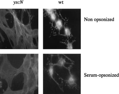
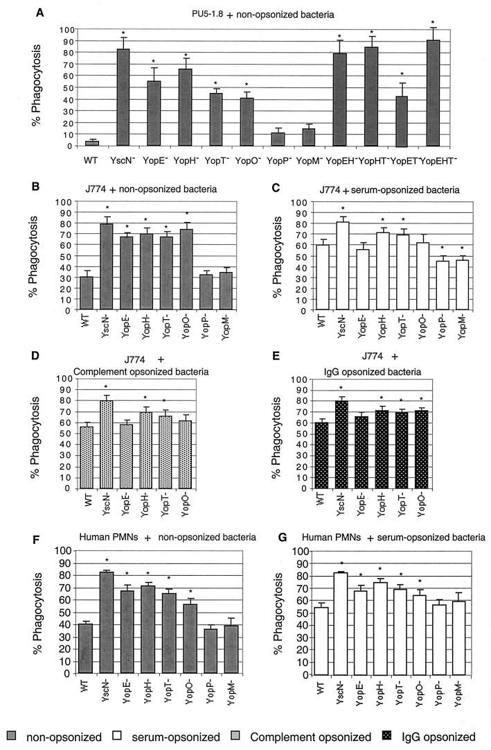
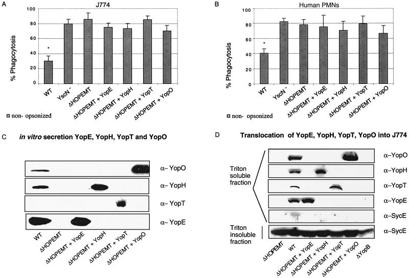
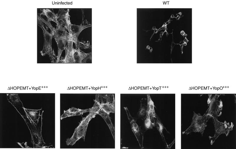
Yersinia enterocolitica is a pathogen endowed with two adhesins, Inv and YadA, and with the Ysc type III secretion system, which allows extracellular adherent bacteria to inject Yop effectors into the cytosol of animal target cells. We tested the influence of all of these virulence determinants on opsonic and nonopsonic phagocytosis by PU5-1.8 and J774 mouse macrophages, as well as by human polymorphonuclear leukocytes (PMNs). The adhesins contributed to phagocytosis in the absence of opsonins but not in the presence of opsonins. In agreement with previous results, YadA counteracted opsonization. In every instance, the Ysc-Yop system conferred a significant level of resistance to phagocytosis. Nonopsonized single-mutant bacteria lacking either YopE, -H, -T, or -O were phagocytosed significantly more by J774 cells and by PMNs. Opsonized bacteria were phagocytosed more than nonopsonized bacteria, and mutant bacteria lacking either YopH, -T, or -O were phagocytosed significantly more by J774 cells and by PMNs than were wild-type (WT) bacteria. Opsonized mutants lacking only YopE were phagocytosed significantly more than were WT bacteria by PMNs but not by J774 cells. Thus, YopH, -T, and -O were involved in all of the phagocytic processes studied here but YopE did not play a clear role in guarding against opsonic phagocytosis by J774. Mutants lacking YopP and YopM were, in every instance, as resistant as WT bacteria. Overexpression of YopE, -H, -T, or -O alone did not confer resistance to phagocytosis, although it affected the cytoskeleton. These results show that YopH, YopT, YopO, and, in some instances, YopE act synergistically to increase the resistance of Y. enterocolitica to phagocytosis by macrophages and PMNs.


Figures

FIG. 1.
FIG. 1.
(A and B) Influence of Inv and YadA on adherence and phagocytosis of Y. enterocolitica MRS40 by J774 macrophages (A and C) and human PMNs (B and D). Nonopsonized or serum-opsonized WT Y. enterocolitica [MRS40(pYV40)], the yscN mutant [MRS40(pMSL41)], the inv mutant [APB40(pYV40)], the yadA mutant [MRS40(pAPB4011)], and the inv yadA mutant [APB40(pAPB4011)] were exposed to J774 cells (A and C) or PMNs (B and D) at a calculated MOI of 50:1 for 30 min at 37°C. The number of adherent bacteria per cell and the percentage of phagocytosis were determined by the double-immunofluorescence technique. Results of adherence are given as the mean plus the standard error of the mean. Phagocytosis results are given as percentages of phagocytosed bacteria relative to the total number of cell-associated bacteria (∗, P < 0.05 compared to the WT; n = 6). The values are the mean plus the standard error of the mean. For each experiment, at least 100 cells were examined.
FIG. 2.
FIG. 2.
Cytotoxic effect of serum-opsonized WT Y. enterocolitica MRS40 bacteria. Rat-1 fibroblasts were infected with Y. enterocolitica [MRS40(pYV40)] bacteria or yscN mutant [MRS40(pMSL41)] bacteria, and cytotoxicity was monitored by staining with FITC-phalloidin after 2 h 30 min of infection. Opsonized and nonopsonized WT Y. enterocolitica MRS40 bacteria were cytotoxic for Rat-1 fibroblasts, in contrast to opsonized or nonopsonized yscN mutant bacteria.
FIG. 3.
FIG. 3.
Phagocytosis of nonopsonized (grey bars, A, B, and F), serum-opsonized (white bars, C and G), complement-opsonized (D), and IgG-opsonized (E) Y. enterocolitica MRS40 and effector mutants by PU5-1.8 macrophages (A), J774 macrophages (B, C, D, and E), and PMNs (F and G). Y. enterocolitica MRS40(pYV40), the yscN mutant [MRS40(pMSL41)], the yopE mutant [MRS40(pAB4052)], the yopH mutant [MRS40(pSI4008)], the yopT mutant [MRS40(pIM409)], the yopO mutant [MRS40(pAB406)], the yopP mutant [MRS40(pMSK41)], the yopM mutant [MRS40(pAB408)], the yopEH mutant [MRS40(pAB404)], the yopHT mutant [MRS40(pIM425)], the yopET mutant [MRS40 (pIM424)], and the yopEHT mutant [MRS40(pIM426)] were exposed to phagocytic cells at a calculated MOI of 50:1 for 30 min at 37°C. Phagocytosis percentages were determined by double-immunofluorescence assay. Phagocytosis results are given as percentages of phagocytosed bacteria relative to the total number of cell-associated bacteria (∗, P < 0.05 compared to the WT; n = 6). The values are the mean plus the standard error of the mean. For each experiment, at least 100 cells were counted.
FIG. 4.
FIG. 4.
(A and B) Phagocytosis of the Y. enterocolitica ΔHOPEMT mutant overexpressing the effectors by J774 macrophages (A) or PMNs (B). J774 macrophages or PMNs were infected at an MOI of 50:1 for 30 min at 37°C with WT [MRS40(pYV40)] Y. enterocolitica, the yscN mutant [MRS4(pMSL41)], the ΔHOPEMT mutant [MRS40(pIML421)], or the ΔHOPEMT strain overexpressing YopE (pMSL68), YopH (pMSL69), YopT (pIML279), or YopO (pMSL34). Phagocytosis percentages were determined by double-immunofluorescence assay. Phagocytosis results are given as percentages of phagocytosed bacteria relative to the total number of cell-associated bacteria (∗, P < 0.05 compared to the ΔHOPEMT strain; n = 6). The values are the mean plus the standard error of the mean. For each experiment, at least 100 cells were counted. (C) Yop secretion by the Y. enterocolitica ΔHOPEMT mutant overexpressing the effectors. Western blot analysis (chemiluminescence detection) of proteins from culture supernatant corresponding to 2 ml of a culture of WT [MRS40(pYV40)] Y. enterocolitica, the yscN mutant [MRS40(pMSL41)], the ΔHOPEMT mutant [MRS40(pIML421)], or the ΔHOPEMT mutant overexpressing YopE (pMSL68), YopH (pMSL69), YopT (pIML279), or YopO (pMSL34) incubated for 4 h at 37°C in BHI-Ox. Immunoblot assay with rabbit anti-YopH, anti-YopT, and anti-YopO polyclonal antibodies and rat anti-YopE monoclonal antibodies (13A9). (D) Injection of the effectors by the various Y. enterocolitica ΔHOPEMT recombinant strains. J774 macrophages were infected at an MOI of 50:1 with WT Y. enterocolitica [MRS40(pYV40)], the ΔHOPEMT mutant [MRS40(pIML421)], the ΔHOPEMT mutant expressing YopE (pMSL68), the ΔHOPEMT mutant expressing YopH (pMSL69), the ΔHOPEMT mutant expressing YopT (pIML279), the ΔHOPEMT mutant expressing YopO (pMSL34), or the yopB mutant [MRS40(pPW401)] producing the same individual Yops. After 2 h, cytosolic fractions of J774 were prepared by Triton (0.1%) lysis and analyzed by SDS-PAGE and Western blotting with rabbit anti-YopH, anti-YopT, anti-YopO, and anti-SycE polyclonal antibodies and rat anti-YopE monoclonal antibodies (13A9). The absence of detection of SycE in the Triton-soluble fraction demonstrates that this fraction does not contain cytoplasmic proteins from Y. enterocolitica.
FIG. 5.
FIG. 5.
Actin disruption by YopE, YopH, YopT, and YopO. Rat-1 cells were infected with Y. enterocolitica MRS40(pYV40) for 2 h 30 min in the presence of 10% FBS. After fixing and permeabilization of the cells, actin was stained with phalloidin-FITC. The cells were then analyzed by confocal scanning laser microscopy. Uninfected cells display elongated actin filaments (stress fibers) throughout the cells. Infection with WT Y. enterocolitica [MRS40(pYV40)], the ΔHOPEMT mutant [MRS40(pIML421)], and the ΔHOPEMT mutant expressing YopE (pMSL68), YopH (pMSL69), YopT (pIML279), or YopO (pMSL34) results in different modifications of the actin cytoskeleton.

Similar articles

Cited by

References

    1. Aderem, A., and D. Underhill. 1999. Mechanisms of phagocytosis in macrophages. Annu. Rev. Immunol. 17:593-623. - PubMed
    1. Allen, L., and A. Aderem. 1996. Molecular definition of distinct cytoskeletal structures involved in complement- and Fc receptor-mediated phagocytosis in macrophages. J. Exp. Med. 184:627-637. - PMC - PubMed
    1. Andersson, K., N. Carballeira, K. Magnusson, C. Persson, O. Stendahl, H. Wolf-Watz, and M. Fallman. 1996. YopH of Yersinia pseudotuberculosis interrupts early phosphotyrosine signalling associated with phagocytosis. Mol. Microbiol. 20:1057-1069. - PubMed
    1. Andersson, K., K. E. Magnusson, M. Majeed, O. Stendahl, and M. Fallman. 1999. Yersinia pseudotuberculosis-induced calcium signaling in neutrophils is blocked by the virulence effector YopH. Infect. Immun. 67:2567-2574. - PMC - PubMed
    1. Andor, A., K. Trulzsch, M. Essler, A. Roggenkamp, A. Wiedemann, J. Heesemann, and M. Aepfelbacher. 2001. YopE of yersinia, a GAP for Rho GTPases, selectively modulates Rac-dependent actin structures in endothelial cells. Cell Microbiol. 3:301-310. - PubMed

Publication types

MeSH terms

LinkOut - more resources