. 2002 Aug;70(8):4204-14.
doi: 10.1128/IAI.70.8.4204-4214.2002.

Yersiniabactin production requires the thioesterase domain of HMWP2 and YbtD, a putative phosphopantetheinylate transferase

Affiliations

Yersiniabactin production requires the thioesterase domain of HMWP2 and YbtD, a putative phosphopantetheinylate transferase

Alexander G Bobrov et al. Infect Immun. 2002 Aug.

Abstract

One requirement for the pathogenesis of Yersinia pestis, the causative agent of bubonic plague, is the yersiniabactin (Ybt) siderophore-dependent iron transport system that is encoded within a high-pathogenicity island (HPI) within the pgm locus of the Y. pestis chromosome. Nine gene products within the HPI have demonstrated functions in the nonribosomal peptide synthesis (NRPS)/polyketide (PK) synthesis or transport of Ybt. NRPS/PK synthetase or synthase enzymes are generally activated by phosphopantetheinylation. However, no products with similarities to known phosphopantetheinyl (P-pant) transferases were found within the pgm locus. We have identified a gene, ybtD, encoded outside the HPI and pgm locus, that is necessary for function of the Ybt system and has similarities to other P-pant transferases such as EntD of Escherichia coli. A deletion within ybtD yielded a strain (KIM6-2085+) defective in siderophore production. This strain was unable to grow on iron-deficient media at 37 degrees C but could be cross-fed by culture supernatants from Ybt-producing strains of Y. pestis. The promoter region of ybtD was fused to lacZ; beta-galactosidase expression from this reporter was not regulated by the iron status of the bacterial cells or by YbtA, a positive regulator of other genes of the ybt system. The ybtD mutant failed to express indicator Ybt proteins (high-molecular-weight protein 1 [HMWP1], HMWP2, and Psn), a pattern similar to those seen with several other ybt biosynthetic mutants. In contrast, cells containing a single amino acid substitution (S2908A) in the terminal thioesterase domain of HMWP2 failed to exhibit any ybt regulatory defects but did not elaborate extracellular Ybt under iron-deficient conditions.


Figures

FIG. 1.
FIG. 1.
Region of Y. pestis KIM10+ genome containing ybtD. The genes encoding two asparaginyl tRNAs, a putative periplasmic binding protein (PBP) for a C4-dicarboxylate ABC transporter, a RafA-like α-galactosidase, and YbtD are indicated as well as IS285 and IS100 elements. Arrows indicate the direction of transcription of selected genes.
FIG. 2.
FIG. 2.
Genetic organization of the Y. pestis ybtD region showing restriction sites used. (A) Dashed line indicates the region deleted in the ΔybtD mutant. The PCR product used in complementation studies is also indicated. The BamHI and XbaI sites in parentheses are artificial restriction sites introduced by PCR. (B) Putative −10 and −35 regions, potential ribosomal binding sites (RBS), and a region with similarity to a Fur binding site (underlined nucleotides), as well as the potential protein start sites, are indicated (underlined and in boldface type). Arrows show the two promoter regions tested in expression studies.
FIG. 3.
FIG. 3.
Amino acid sequence alignment of YbtD from Y. pestis, NgrA from P. luminescens, VibD from V. cholerae, and EntD from E. coli. Residues with identity to YbtD are in white with a solid black background. Conservative and semiconservative amino acid substitutions are shaded. The consensus line shows identical residues in all four proteins (uppercase letters) and identical residues in two or more proteins (lowercase letters). The identity and similarity of YbtD to each of these proteins are 34 and 65.3% (NgrA), 31.3 and 60.2% (VibD), and 27.2 and 58.5% (EntD). The residues below the consensus line indicate conserved (lowercase) and highly conserved (uppercase) amino acids within the proposed P-pant transferase domain derived from comparison of 22 P-pant transferases (46).
FIG. 4.
FIG. 4.
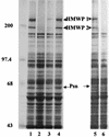
SDS-PAGE analysis of whole-cell proteins from Y. pestis strains grown in iron-deficient PMH2. Cultures from Y. pestis KIM6+ (lane 1), KIM6-2046.1 (irp2::kan2046.1) (lane 2), KIM6-2085+ (ΔybtD2085) (lane 3), and KIM6-2085(pYBTD4)+ (ΔybtD2085/ybtD+) (lane 4) were incubated with 35S-labeled amino acids for 1 h. To demonstrate the effect of exogenous siderophore on expression of proteins by KIM6-2085+ cells, KIM6+ culture supernatant containing Ybt siderophore (lane 5) or KIM6-2046.1 culture supernatant (lane 6) was added 1:1 at the same time as 35S-labeled amino acids. Total cellular proteins were separated on a 9% polyacrylamide gel and visualized by autoradiography. Sizes of molecular mass markers (in kilodaltons) are indicated. Arrows point to the iron-regulated proteins HMWP1 (240 kDa), HMWP2 (190 kDa), and Psn (68 kDa).
FIG. 5.
FIG. 5.
Conserved TE domains of YbtT and HMWP2. The TE consensus sequence is described in reference . The serine residue in HMWP1 that was changed to an alanine in KIM6-2086 is underlined.
FIG. 6.
FIG. 6.
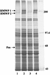
SDS-PAGE analysis of whole-cell proteins from Y. pestis strains grown in iron-sufficient and iron-deficient PMH2. Cultures from Y. pestis KIM6+ (lanes 1 and 2), KIM6-2046.1 (irp2::kan2046.1) (lane 3), and KIM6-2086 (irp1-2086) (lane 4) were incubated with 35S-labeled amino acids for 1 h. Total cellular proteins were separated on a 9% polyacrylamide gel and visualized by autoradiography. Cell extracts from iron-deficient cultures (lanes 2 to 4) or iron-sufficient cultures (lane 1) are shown. Sizes of molecular mass markers (in kilodaltons) are indicated. Arrows point to the iron-regulated proteins HMWP1 (240 kDa), HMWP2 (190 kDa), and Psn (68 kDa).

Similar articles

Cited by

References

    1. Armstrong, S. K., G. S. Pettis, L. J. Forrester, and M. A. McIntosh. 1989. The Escherichia coli enterobactin biosynthesis gene, entD: nucleotide sequence and membrane localization of its protein product. Mol. Microbiol. 3:757-766. - PubMed
    1. Ausubel, F. M., R. Brent, R. E. Kingston, D. D. Moore, J. G. Seidman, J. A. Smith, and K. Struhl (ed.). 1987. Current protocols in molecular biology. John Wiley & Sons, New York, N.Y.
    1. Bach, S., C. Buchrieser, M. Prentice, A. Guiyoule, T. Msadek, and E. Carniel. 1999. The high-pathogenicity island of Yersinia enterocolitica Ye8081 undergoes low-frequency deletion but not precise excision, suggesting recent stabilization in the genome. Infect. Immun. 67:5091-5099. - PMC - PubMed
    1. Bearden, S. W., J. D. Fetherston, and R. D. Perry. 1997. Genetic organization of the yersiniabactin biosynthetic region and construction of avirulent mutants in Yersinia pestis. Infect. Immun. 65:1659-1668. - PMC - PubMed
    1. Bearden, S. W., and R. D. Perry. 1999. The Yfe system of Yersinia pestis transports iron and manganese and is required for full virulence of plague. Mol. Microbiol. 32:403-414. - PubMed

Publication types

MeSH terms