

Pathogenesis of pneumococcal pneumonia in cyclophosphamide-induced leukopenia in mice
- PMID: 12117931
- PMCID: PMC128150
- DOI: 10.1128/IAI.70.8.4226-4238.2002
Pathogenesis of pneumococcal pneumonia in cyclophosphamide-induced leukopenia in mice
Abstract
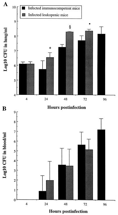
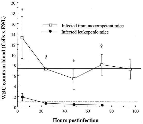
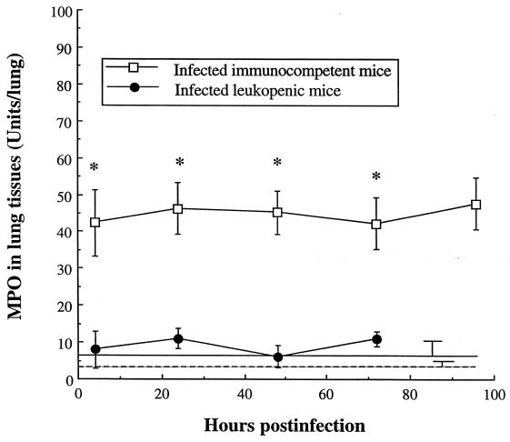
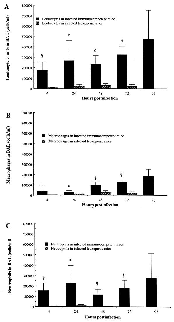
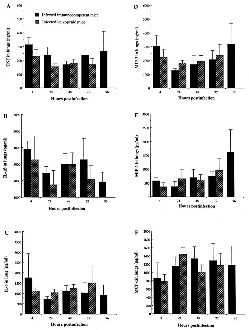
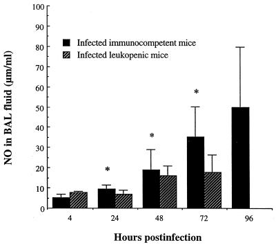
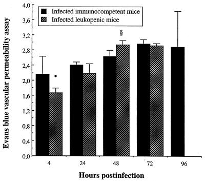
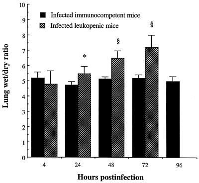
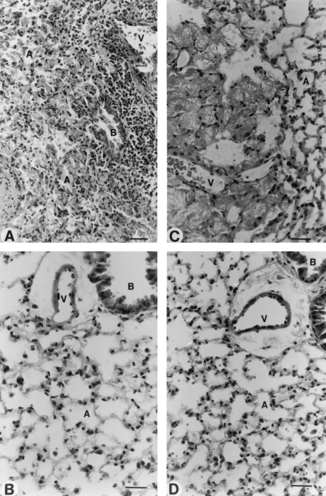
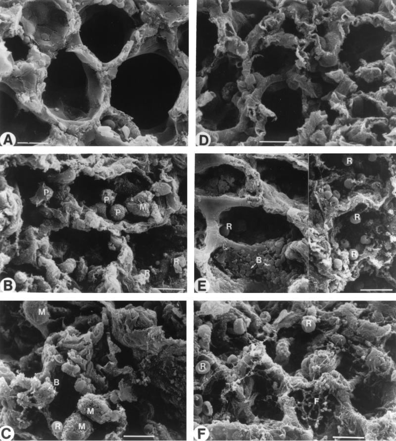
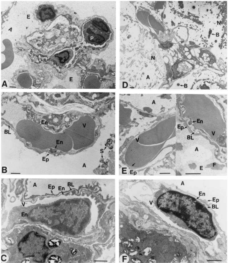
Streptococcus pneumoniae pneumonia frequently occurs in leukopenic hosts, and most patients subsequently develop lung injury and septicemia. However, few correlations have been made so far between microbial growth, inflammation, and histopathology of pneumonia in specific leukopenic states. In the present study, the pathogenesis of pneumococcal pneumonia was investigated in mice rendered leukopenic by the immunosuppressor antineoplastic drug cyclophosphamide. Compared to the immunocompetent state, cyclophosphamide-induced leukopenia did not hamper interleukin-1 (IL-1), IL-6, macrophage inflammatory protein-1 (MIP-1), MIP-2, and monocyte chemotactic protein-1 secretion in infected lungs. Leukopenia did not facilitate bacterial dissemination into the bloodstream despite enhanced bacterial proliferation into lung tissues. Pulmonary capillary permeability and edema as well as lung injury were enhanced in leukopenic mice despite the absence of neutrophilic and monocytic infiltration into their lungs, suggesting an important role for bacterial virulence factors and making obvious the fact that neutrophils are ultimately not required for lung injury in this model. Scanning and transmission electron microscopy revealed extensive disruption of alveolar epithelium and a defect in surfactant production, which were associated with alveolar collapse, hemorrhage, and fibrin deposits in alveoli. These results contrast with those observed in immunocompetent animals and indicate that leukopenic hosts suffering from pneumococcal pneumonia are at a higher risk of developing diffuse alveolar damage.
Figures
Similar articles
-
Microbiological and inflammatory factors associated with the development of pneumococcal pneumonia.J Infect Dis. 2001 Aug 1;184(3):292-300. doi: 10.1086/322021. Epub 2001 Jun 26. J Infect Dis. 2001. PMID: 11443554
-
Role of chemokines and formyl peptides in pneumococcal pneumonia-induced monocyte/macrophage recruitment.J Immunol. 2001 Jun 15;166(12):7353-61. doi: 10.4049/jimmunol.166.12.7353. J Immunol. 2001. PMID: 11390486
-
Pulmonary and systemic host response to Streptococcus pneumoniae and Klebsiella pneumoniae bacteremia in normal and immunosuppressed mice.Infect Immun. 2001 Sep;69(9):5294-304. doi: 10.1128/IAI.69.9.5294-5304.2001. Infect Immun. 2001. PMID: 11500398 Free PMC article.
-
Trivalent pneumococcal protein recombinant vaccine protects against lethal Streptococcus pneumoniae pneumonia and correlates with phagocytosis by neutrophils during early pathogenesis.Vaccine. 2015 Feb 18;33(8):993-1000. doi: 10.1016/j.vaccine.2015.01.014. Epub 2015 Jan 15. Vaccine. 2015. PMID: 25597944
-
Macrophage inflammatory proteins: biology and role in pulmonary inflammation.Exp Lung Res. 1994 Nov-Dec;20(6):473-90. doi: 10.3109/01902149409031733. Exp Lung Res. 1994. PMID: 7882902 Review.
Cited by
-
Biomarkers of thrombosis, fibrinolysis, and inflammation in patients with severe sepsis due to community-acquired pneumonia with and without Streptococcus pneumoniae.Infection. 2009 Aug;37(4):358-64. doi: 10.1007/s15010-008-8128-6. Epub 2009 Jan 23. Infection. 2009. PMID: 19169631 Clinical Trial.
-
Altered pulmonary capillary permeability in immunosuppressed guinea pigs infected with Legionella pneumophila serogroup 1.Exp Ther Med. 2019 Dec;18(6):4368-4378. doi: 10.3892/etm.2019.8102. Epub 2019 Oct 14. Exp Ther Med. 2019. PMID: 31772633 Free PMC article.
-
CXCR3 and its ligands participate in the host response to Bordetella bronchiseptica infection of the mouse respiratory tract but are not required for clearance of bacteria from the lung.Infect Immun. 2005 Jan;73(1):485-93. doi: 10.1128/IAI.73.1.485-493.2005. Infect Immun. 2005. PMID: 15618188 Free PMC article.
-
Differential contribution of bacterial N-formyl-methionyl-leucyl- phenylalanine and host-derived CXC chemokines to neutrophil infiltration into pulmonary alveoli during murine pneumococcal pneumonia.Infect Immun. 2007 Nov;75(11):5361-7. doi: 10.1128/IAI.02008-06. Epub 2007 Aug 20. Infect Immun. 2007. PMID: 17709413 Free PMC article.
-
Organ Failure Due to Systemic Injury in Acute Pancreatitis.Gastroenterology. 2019 May;156(7):2008-2023. doi: 10.1053/j.gastro.2018.12.041. Epub 2019 Feb 12. Gastroenterology. 2019. PMID: 30768987 Free PMC article. Review.
References
-
- Anonymous. 1987. Adult respiratory distress syndrome and neutropenia. N. Engl. J. Med. 316:413-414. - PubMed
-
- Arfors, K. E., C. Lundberg, L. Lindbom, K. Lundberg, P. G. Beatty, and J. M. Harlan. 1987. A monoclonal antibody to the membrane glycoprotein complex CD18 inhibits polymorphonuclear leukocyte accumulation and plasma leakage in vivo. Blood 69:338-340. - PubMed
-
- Austrian, R., and J. Gold. 1964. Pneumococcal bacteremia with especial reference to bacteremic pneumococcal pneumonia. Ann. Intern. Med. 60:759-776. - PubMed
-
- Bachofen, A., and E. R. Weibel. 1977. Alterations of the gas exchange apparatus in adult respiratory insufficiency associated with septicemia. Am. Rev. Respir. Dis. 116:589-615. - PubMed
-
- Baird, B. R., J. C. Cheronis, R. A. Sandhaus, E. M. Berger, C. W. White, and J. E. Repine. 1986. O2 metabolites and neutrophil elastase synergistically cause edematous injury in isolated rat lungs. J. Appl. Physiol 61:2224-2229. - PubMed
MeSH terms
Substances
LinkOut - more resources
Full Text Sources
Research Materials
