

Induction of cell-mediated immunity to Staphylococcus aureus in the mouse mammary gland by local immunization with a live attenuated mutant
- PMID: 12117934
- PMCID: PMC128206
- DOI: 10.1128/IAI.70.8.4254-4260.2002
Induction of cell-mediated immunity to Staphylococcus aureus in the mouse mammary gland by local immunization with a live attenuated mutant
Abstract
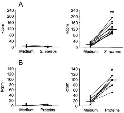
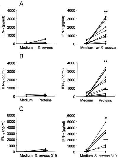
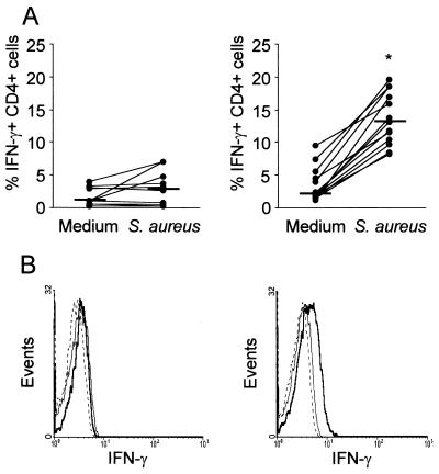
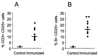
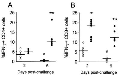
The efficacy of intramammary (Ima) immunization with a live attenuated (la) Staphylococcus aureus mutant to protect the mouse mammary gland from infection has previously been established. The present study was aimed at evaluating whether Ima immunization with la-S. aureus can induce cell-mediated immune responses to the pathogen within the mammary gland. Mice were immunized by Ima route with la-S. aureus, and regional lymph node mononuclear cells were obtained thereafter. A higher expression of the interleukin-2 receptor was found on B and T cells from immunized mice when they were compared with control mice. Immunization with la-S. aureus induced strong proliferative responses to S. aureus. Moreover, significantly increased levels of gamma interferon (IFN-gamma) were produced by CD4+ T cells when lymphocytes from immunized mice, but not from control mice, were cultured in the presence of staphylococcal antigens. Moreover, a significant increase in the percentage of IFN-gamma-producing CD4+ and CD8+ T cells was observed after S. aureus Ima challenge in immunized mice compared to challenged control mice. Our results demonstrated that Ima immunization with la-S. aureus induced primed lymphocyte populations capable of responding against staphylococcal antigens during in vitro stimulation, as well as during in vivo infection by S. aureus. CD4+ and CD8+ T cells appear to be the main lymphocyte subpopulations involved in this response. It is suggested that IFN-gamma production induced by Ima immunization may play a pivotal role in the eradication of intracellular staphylococci.
Figures
Similar articles
-
CD8α- conventional dendritic cells control Vβ T-cell immunity in response to Staphylococcus aureus infection in mice.Immunology. 2020 Apr;159(4):404-412. doi: 10.1111/imm.13171. Epub 2020 Jan 21. Immunology. 2020. PMID: 31909831 Free PMC article.
-
Mechanisms of fibronectin-binding protein A (FnBPA110-263) vaccine efficacy in Staphylococcus aureus sepsis versus skin infection.Clin Immunol. 2018 Sep;194:1-8. doi: 10.1016/j.clim.2018.05.007. Epub 2018 Jun 12. Clin Immunol. 2018. PMID: 29906512
-
A mutant of staphylococcal enterotoxin C devoid of bacterial superantigenic activity elicits a Th2 immune response for protection against Staphylococcus aureus infection.Infect Immun. 2005 Jan;73(1):174-80. doi: 10.1128/IAI.73.1.174-180.2005. Infect Immun. 2005. PMID: 15618152 Free PMC article.
-
Vaccination with a live-attenuated small-colony variant improves the humoral and cell-mediated responses against Staphylococcus aureus.PLoS One. 2019 Dec 27;14(12):e0227109. doi: 10.1371/journal.pone.0227109. eCollection 2019. PLoS One. 2019. PMID: 31881064 Free PMC article.
-
Adaptive Cell-Mediated Immunity in the Mammary Gland of Dairy Ruminants.Front Vet Sci. 2022 Apr 5;9:854890. doi: 10.3389/fvets.2022.854890. eCollection 2022. Front Vet Sci. 2022. PMID: 35464360 Free PMC article. Review.
Cited by
-
Suppression of the inflammatory immune response prevents the development of chronic biofilm infection due to methicillin-resistant Staphylococcus aureus.Infect Immun. 2011 Dec;79(12):5010-8. doi: 10.1128/IAI.05571-11. Epub 2011 Sep 26. Infect Immun. 2011. PMID: 21947772 Free PMC article.
-
Efficacy of Aloe vera, Ananas comosus, and Sansevieria masoniana Cream on the Skin Wound Infected with MRSA.Adv Pharmacol Sci. 2018 Apr 19;2018:4670569. doi: 10.1155/2018/4670569. eCollection 2018. Adv Pharmacol Sci. 2018. PMID: 29849604 Free PMC article.
-
Vitexin Mitigates Staphylococcus aureus-Induced Mastitis via Regulation of ROS/ER Stress/NF-κB/MAPK Pathway.Oxid Med Cell Longev. 2022 Jun 27;2022:7977433. doi: 10.1155/2022/7977433. eCollection 2022. Oxid Med Cell Longev. 2022. PMID: 35795861 Free PMC article.
-
CD4 T cell antigens from Staphylococcus aureus Newman strain identified following immunization with heat-killed bacteria.Clin Vaccine Immunol. 2012 Apr;19(4):477-89. doi: 10.1128/CVI.05642-11. Epub 2012 Feb 8. Clin Vaccine Immunol. 2012. PMID: 22323557 Free PMC article.
-
Phytol-based novel adjuvants in vaccine formulation: 2. Assessment of efficacy in the induction of protective immune responses to lethal bacterial infections in mice.J Immune Based Ther Vaccines. 2006 Oct 23;4:5. doi: 10.1186/1476-8518-4-5. J Immune Based Ther Vaccines. 2006. PMID: 17059608 Free PMC article.
References
-
- Bancroft, G. J., R. D. Schreiber, and E. R. Unanue. 1991. Natural immunity: a T-cell-independent pathway of macrophage activation defined in the scid mouse. Immunol. Rev. 124:5-24. - PubMed
-
- Ferens, W. A., and G. A. Bohach. 2000. Persistence of Staphylococcus aureus on mucosal membranes: superantigens and internalization by host cells. J. Lab. Clin. Med. 135:225-230. - PubMed
-
- Foster, T. J. 1991. Potential for vaccination against infections caused by Staphylococcus aureus. Vaccine 9:221-227. - PubMed
-
- Fox, L. K., H. D. Liggit, T. Yilma, and L. B. Corbeil. 1990. The effects of interferon intramammary administration on mammary phagocyte function. Zentbl. Veterinarmed. [B] 37:28-30. - PubMed
Publication types
MeSH terms
Substances
LinkOut - more resources
Full Text Sources
Medical
Research Materials
