

Autotransported serine protease A of Neisseria meningitidis: an immunogenic, surface-exposed outer membrane, and secreted protein
- PMID: 12117956
- PMCID: PMC128147
- DOI: 10.1128/IAI.70.8.4447-4461.2002
Autotransported serine protease A of Neisseria meningitidis: an immunogenic, surface-exposed outer membrane, and secreted protein
Abstract
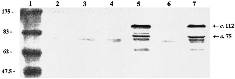
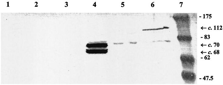
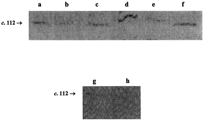
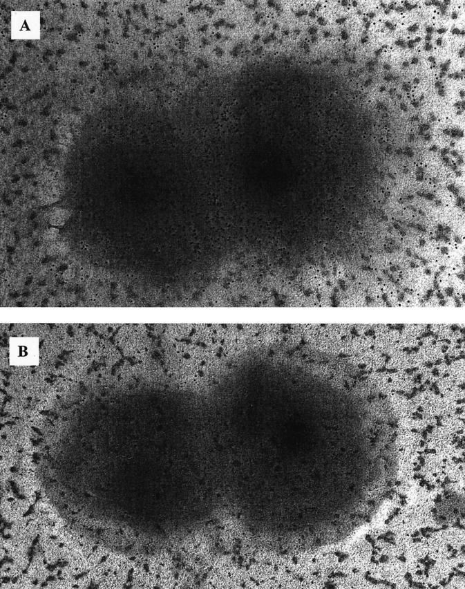
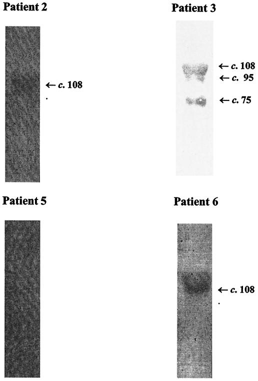
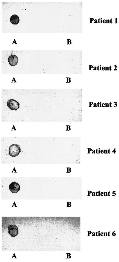
Several autotransporter proteins have previously been identified in Neisseria meningitidis. Using molecular features common to most members of the autotransporter family of proteins, we have identified an additional novel ca. 112-kDa autotransporter protein in the meningococcal genomic sequence data. This protein, designated autotransported serine protease A (AspA), has significant N-terminal homology to the secreted serine proteases (subtilases) from several organisms and contains a serine protease catalytic triad. The amino acid sequence of AspA is well-conserved in serogroup A, B, and C meningococci. In Neisseria gonorrhoeae, the AspA homologue appears to be a pseudogene. The gene encoding AspA was cloned and expressed from meningococcal strain MC58 (B15:P1.16b). Anti-AspA antibodies were detected in patients' convalescent-phase sera, suggesting that AspA is expressed in vivo during infection and is immunogenic and cross-reactive. Rabbit polyclonal monospecific anti-AspA serum was used to probe whole-cell proteins from a panel of wild-type meningococcal strains and two AspA mutant strains. Expression of the ca. 112-kDa precursor polypeptide was detected in 12 of 20 wild-type meningococcal strains examined, suggesting that AspA expression is phase variable. Immunogold electron microscopy and cellular fractionation studies showed that the AspA precursor is transported to the outer membrane and remains surface exposed. Western blot experiments confirmed that smaller, ca. 68- or 70-kDa components of AspA (AspA68 and AspA70, respectively) are then secreted into the meningococcal culture supernatant. Site-directed mutagenesis of S426 abolished secretion of both rAspA68 and rAspA70 in Escherichia coli, confirming that AspA is an autocleaved autotransporter protein. In conclusion, we characterized a novel, surface-exposed and secreted, immunogenic, meningococcal autotransporter protein.
Figures

Similar articles
-
Identification and characterization of App: an immunogenic autotransporter protein of Neisseria meningitidis.Mol Microbiol. 2001 Aug;41(3):611-23. doi: 10.1046/j.1365-2958.2001.02516.x. Mol Microbiol. 2001. PMID: 11532129
-
Genetic Similarity of Gonococcal Homologs to Meningococcal Outer Membrane Proteins of Serogroup B Vaccine.mBio. 2019 Sep 10;10(5):e01668-19. doi: 10.1128/mBio.01668-19. mBio. 2019. PMID: 31506309 Free PMC article.
-
Characterization of MspA, an immunogenic autotransporter protein that mediates adhesion to epithelial and endothelial cells in Neisseria meningitidis.Infect Immun. 2006 May;74(5):2957-64. doi: 10.1128/IAI.74.5.2957-2964.2006. Infect Immun. 2006. PMID: 16622234 Free PMC article.
-
The autodisplay story, from discovery to biotechnical and biomedical applications.Microbiol Mol Biol Rev. 2007 Dec;71(4):600-19. doi: 10.1128/MMBR.00011-07. Microbiol Mol Biol Rev. 2007. PMID: 18063719 Free PMC article. Review.
-
Genetic mechanisms and biological implications of phase variation in pathogenic neisseriae.Clin Microbiol Rev. 1989 Apr;2 Suppl(Suppl):S139-45. doi: 10.1128/CMR.2.Suppl.S139. Clin Microbiol Rev. 1989. PMID: 2655883 Free PMC article. Review. No abstract available.
Cited by
-
Functional characterization of AasP, a maturation protease autotransporter protein of Actinobacillus pleuropneumoniae.Infect Immun. 2008 Dec;76(12):5608-14. doi: 10.1128/IAI.00085-08. Epub 2008 Oct 13. Infect Immun. 2008. PMID: 18852244 Free PMC article.
-
Characterization and Distribution of the autB Gene in Neisseria meningitidis.Front Cell Infect Microbiol. 2017 Oct 6;7:436. doi: 10.3389/fcimb.2017.00436. eCollection 2017. Front Cell Infect Microbiol. 2017. PMID: 29057217 Free PMC article.
-
Comparative analysis of the biochemical and functional properties of C-terminal domains of autotransporters.J Bacteriol. 2010 Nov;192(21):5588-602. doi: 10.1128/JB.00432-10. Epub 2010 Aug 27. J Bacteriol. 2010. PMID: 20802036 Free PMC article.
-
The Serine Protease Autotransporters TagB, TagC, and Sha from Extraintestinal Pathogenic Escherichia coli Are Internalized by Human Bladder Epithelial Cells and Cause Actin Cytoskeletal Disruption.Int J Mol Sci. 2020 Apr 26;21(9):3047. doi: 10.3390/ijms21093047. Int J Mol Sci. 2020. PMID: 32357479 Free PMC article.
-
The AprV5 subtilase is required for the optimal processing of all three extracellular serine proteases from Dichelobacter nodosus.PLoS One. 2012;7(10):e47932. doi: 10.1371/journal.pone.0047932. Epub 2012 Oct 24. PLoS One. 2012. PMID: 23112874 Free PMC article.
References
-
- Abdel-Hadi, H., K. G. Wooldridge, K. Robinson, and D. A. A. Ala'Aldeen. 2001. Identification and characterization of App: an immunogenic autotransporter protein of Neisseria meningitidis. Mol. Microbiol. 41:611-623. - PubMed
-
- Ait-Tahar, K., K. G. Wooldridge, D. P. J. Turner, M. Atta, I. Todd, and D. A. A. Ala'Aldeen. 2000. Autotransporter A protein of Neisseria meningitidis: a potent CD4+ T-cell and B-cell stimulating antigen detected by expression cloning. Mol. Microbiol. 37:1094-1105. - PubMed
-
- Barenkamp, S. J., and J. W. St. Geme III. 1996. Identification of a second family of high-molecular-weight adhesion proteins expressed by non-typable Haemophilus influenzae. Mol. Microbiol. 19:1215-1223. - PubMed
Publication types
MeSH terms
Substances
Associated data
- Actions
- Actions
LinkOut - more resources
Full Text Sources
Other Literature Sources
