

Sequential expression of the neuropeptides substance P and somatostatin in granulomas associated with murine cysticercosis
- PMID: 12117965
- PMCID: PMC128166
- DOI: 10.1128/IAI.70.8.4534-4538.2002
Sequential expression of the neuropeptides substance P and somatostatin in granulomas associated with murine cysticercosis
Abstract
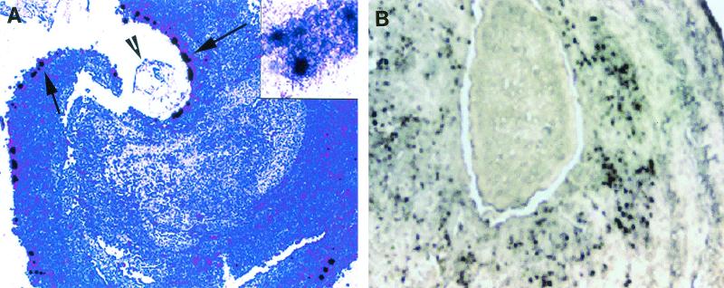
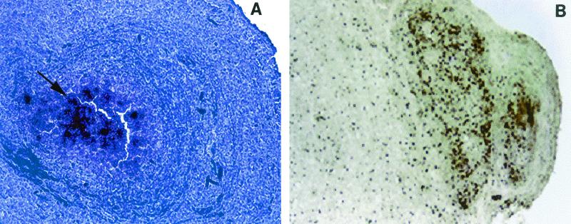
Neurocysticercosis, a parasitic infection of the human central nervous system caused by Taenia solium, is a leading cause of seizures. Seizures associated with neurocysticercosis are caused mainly by the host inflammatory responses to dying parasites in the brain parenchyma. We previously demonstrated sequential expression of Th1 cytokines in early-stage granulomas, followed by expression of Th2 cytokines in later-stage granulomas in murine cysticercosis. However, the mechanism leading to this shift in cytokine response in the granulomas is unknown. Neuropeptides modulate cytokine responses and granuloma formation in murine schistosomiasis. Substance P (SP) induces Th1 cytokine expression and granuloma formation, whereas somatostatin inhibits the granulomatous response. We hypothesized that neuropeptides might play a role in regulation of the granulomatous response in cysticercosis. To test this hypothesis, we compared expression of SP and expression of somatostatin in murine cysticercal granulomas by using in situ hybridization and immunohistochemistry. We also compared expression with granuloma stage. Expression of SP mRNA was more frequent in the early-stage granulomas than in the late-stage granulomas (34 of 35 early-stage granulomas versus 1 of 13 late-stage granulomas). By contrast, somatostatin was expressed primarily in later-stage granulomas (13 of 14 late-stage granulomas versus 2 of 35 early-stage granulomas). The median light microscope grade of SP mRNA expression in the early-stage granulomas was significantly higher than that in the late-stage granulomas (P = 0.008, as determined by the Wilcoxon signed rank test). By contrast, somatostatin mRNA expression was higher at later stages (P = 0.008, as determined by the Wilcoxon signed rank test). SP and somatostatin are therefore temporally expressed in granulomas associated with murine cysticercosis, which may be related to differential expression of Th1 and Th2 cytokines.
Figures
References
-
- Berczi, I., I. M. Chalmers, E. Nagy, and R. J. Warrington. 1996. The immune effects of neuropeptides. Baillieres Clin. Rheumatol. 10:227-257. - PubMed
-
- Blum, A. M., D. E. Elliott, A. Metwali, J. Li, K. Qadir, and J. V. Weinstock. 1998. Substance P regulates somatostatin expression in inflammation. J. Immunol. 161:6316-6322. - PubMed
-
- Blum, A. M., A. Metwali, M. Kim-Miller, J. Li, K. Qadir, D. E. Elliott, B. Lu, Z. Fabry, N. Gerard, and J. V. Weinstock. 1999. The substance P receptor is necessary for a normal granulomatous response in murine schistosomiasis mansoni. J. Immunol. 162:6080-6085. - PubMed
-
- Blum, A. M., A. Metwali, R. C. Mathew, G. Cook, D. Elliott, and J. V. Weinstock. 1992. Granuloma T lymphocytes in murine schistosomiasis mansoni have somatostatin receptors and respond to somatostatin with decreased IFN-gamma secretion. J. Immunol. 149:3621-3626. - PubMed
-
- Cook, G. A., D. Elliott, A. Metwali, A. M. Blum, M. Sandor, R. Lynch, and J. V. Weinstock. 1994. Molecular evidence that granuloma T lymphocytes in murine schistosomiasis mansoni express an authentic substance P (NK-1) receptor. J. Immunol. 152:1830-1835. - PubMed
Publication types
MeSH terms
Substances
Grants and funding
LinkOut - more resources
Full Text Sources
