. 2002 Aug;70(8):4571-80.
doi: 10.1128/IAI.70.8.4571-4580.2002.

Opsonization modulates Rac-1 activation during cell entry by Leishmania amazonensis

Affiliations

Opsonization modulates Rac-1 activation during cell entry by Leishmania amazonensis

J Morehead et al. Infect Immun. 2002 Aug.

Abstract

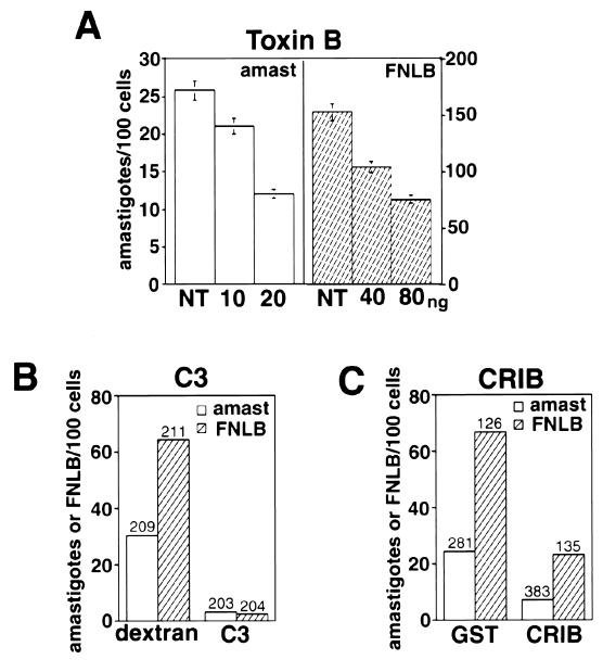
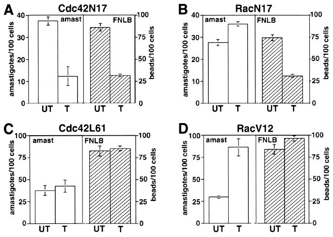
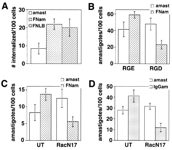
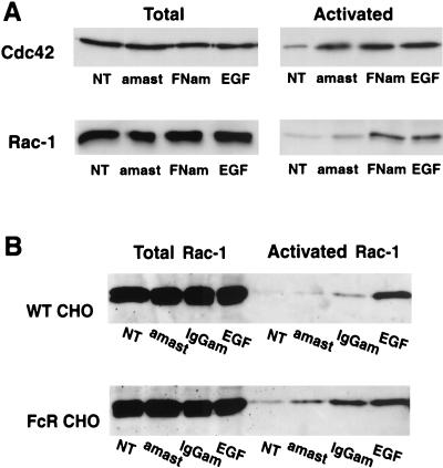
Lesions caused by Leishmania amazonensis normally heal, but relapses occur due to parasite persistence in host tissues. It has been proposed that infection of fibroblasts plays an important role in this process by providing the parasites with a safe haven in which to replicate. However, most previous studies have focused on the entry of Leishmania into macrophages, a process mediated by serum opsonins. To gain insight into a possible role of nonopsonic entry in the intracellular persistence of amastigotes, we examined the invasion of Chinese hamster ovary (CHO) cells. Amastigotes entered CHO cells by a cytochalasin D, genistein, wortmannin, and 2,3-butanedione monoxime-sensitive pathway and replicated within phagolysosomes. However, unlike most phagocytic processes described to date, amastigote internalization in CHO cells involved activation of the GTPases Rho and Cdc42 but not Rac-1. When uptake was mediated by fibronectin or when amastigotes were opsonized with immunoglobulin G and internalized by Fc receptor-expressing CHO cells, Rac-1 activation was restored and found to be required for parasite internalization. Given the essential role of Rac in assembly of the respiratory burst oxidase, invasion through this nonopsonic, Rac-1-independent pathway may play a central role in the intracellular survival of Leishmania in immune hosts.


Figures

FIG. 1.
FIG. 1.
L. amazonensis amastigotes enter CHO cells by phagocytosis. CHO cells were exposed to 5 × 10⁷ amastigotes/ml for 20 min, fixed, and processed for TEM. (A) Pseudopod formation at the amastigote attachment site. (B) Partially formed phagosome. (C) Complete phagosome. Arrowheads point to pseudopod extensions, and arrows point to the membrane of a recently formed phagosome. P, parasite. Sizing bars, 1 μm. (D) Digital fluorescence microscopy image of a CHO cell containing one internalized amastigote (long arrow) and with two amastigotes in the process of being phagocytosed (short arrow). The cells were fixed and stained with rhodamine-phalloidin (to reveal the cups of polymerized actin surrounding the parasites), DAPI (to reveal the CHO and amastigote nuclei), and fluorescein isothiocyanate-labeled anti-L. amazonensis MAb (to reveal extracellular portions of the parasites). (E) Rhodamine-phalloidin staining of an infected CHO cell, showing the F-actin cup surrounding an amastigote (arrowhead). (F) Same cell (shown in panel E) stained with anti-vinculin antibodies, showing vinculin associated with the phagocytic cup (arrowhead).
FIG. 2.
FIG. 2.
Agents that block phagocytosis inhibit internalization of FNLB and L. amazonensis amastigotes in CHO cells. NT, not treated. (A) Number of intracellular parasites in cells not treated or treated with 10 μM CD. (B) Number of intracellular parasites in cells not treated or treated with 25 mM BDM or 20 μM GT. (C) Number of intracellular parasites in cells not treated or treated with 100 nM WM or 100 μM LY. CHO cells were pretreated with the indicated drugs for 20 min, followed by exposure to amastigotes for 60 min. (D) TEM image of a CHO cell containing an amastigote and an FNLB. CHO cells were simultaneously exposed to FNLB and amastigotes for 60 min. P, parasite; B, FNLB. Sizing bar, 1 μm. (E) Number of intracellular FNLB in CHO cells not treated or treated with BDM or GT as described for panel B. (F) Number of intracellular FNLB in CHO cells not treated or treated with WM or LY as described for panel C. CHO cells were pretreated with the indicated drugs for 20 min, followed by exposure to FNLB for 60 min. The data represent the means and standard deviations (SD) of triplicate experiments.
FIG. 3.
FIG. 3.
L. amazonensis replicates within phagolysosomes in CHO cells. (A) Phase-contrast image (top) and immunofluorescence staining with anti-LAMP-1 MAbs (bottom) of CHO cells infected with L. amazonensis amastigotes. Arrows point to intracellular parasites, and arrowheads point to extracellularly attached parasites. (B) Phagolysosome containing replicating amastigotes at 24 (top) and 48 (bottom) h after infection. (C) TEM showing amastigotes replicating in a phagolysosome containing LDL-gold particles 24 h after infection. Arrowheads point to LDL-gold particles in the lumen of the phagolysosome, the white arrow points to LDL-gold particles internalized by one of the parasites, and the black arrow points to the phagolysosome membrane. P, parasite. Sizing bar, 1 μm. (D) Intracellular growth curve of L. amazonensis in CHO cells. Cells were exposed to amastigotes for 60 min, washed, and further incubated at 34°C for 24 and 48 h. After fixation, the total number of intracellular parasites per five microscopic fields (×100) was determined in triplicate (the data represent the averages ± SD of triplicate experiments).
FIG. 4.
FIG. 4.
Nonopsonic cell entry by L. amazonensis requires activation of Rho GTPases. (A) Number of intracellular amastigotes (amast) or FNLB in cells not treated (NT) or treated with the indicated amounts of toxin B/ml for 60 min. After microinjection, CHO cells were exposed to amastigotes or FNLB for 60 min at 34°C and fixed and the number of internalized particles was determined in triplicate (the data correspond to the means and SD of triplicate experiments). (B) Number of intracellular amastigotes or FNLB in cells microinjected with dextran alone or with C3 exotoxin. (C) Number of intracellular amastigotes or FNLB in cells microinjected with GST alone or with CRIB-GST. After microinjection, CHO cells were exposed to amastigotes or FNLB for 60 min at 34°C and fixed and the number of internalized particles was determined in the microinjected cells (the numbers above the columns indicate the total number of microinjected cells analyzed).
FIG. 5.
FIG. 5.
Nonopsonic cell entry by L. amazonensis requires activation of Cdc42 but not of Rac-1. (A) Number of intracellular amastigotes or FNLB in cells not transfected (UT) or transfected with Cdc42N17. (B) Number of intracellular amastigotes or FNLB in cells not transfected (UT) or transfected with RacN17. (C) Number of intracellular amastigotes or FNLB in cells not transfected (UT) or transfected with Cdc42L61. (D) Number of intracellular amastigotes or FNLB in cells not transfected (UT) or transfected with RacV12. Transfected CHO cells were exposed to amastigotes or FNLB for 60 min at 34°C and fixed, and the number of internalized particles was determined in triplicate (the data correspond to the means and SD of triplicate experiments).
FIG. 6.
FIG. 6.
Rac-1 activation is required for opsonin-mediated uptake of L. amazonensis. (A) Number of intracellular amastigotes (amast), fibronectin-coated amastigotes (FNam), or FNLB in CHO cells incubated at 4°C with amastigotes or FNLB for 30 min, followed by incubation at 34°C for 30 min. (B) Number of amastigotes or fibronectin-coated amastigotes internalized in the presence of 100 μM RGE or RGD peptides (Life Technologies). CHO cells were exposed to amastigotes for 60 min at 34°C. (C) Number of amastigotes or fibronectin-coated amastigotes internalized in cells not transfected (UT) or transfected with RacN17. Transfected CHO cells were exposed to amastigotes for 60 min at 34°C. (D) Number of amastigotes or IgG-coated amastigotes (IgGam) internalized in FcRII-B2 cells not transfected (UT) or transfected with RacN17. FcRII-B2-transfected CHO cells were exposed to amastigotes for 60 min at 34°C. After fixation, the number of internalized particles was determined in triplicate (the data correspond to the means and SD of triplicate experiments).
FIG. 7.
FIG. 7.
Nonopsonic cell entry by L. amazonensis does not trigger Rac-1 activation. The total endogenous levels of Cdc42 and Rac-1 were determined by Western blotting in lysates of CHO cells. The levels of activated (GTP-bound) forms of the same GTPases were determined according to their ability to bind GST-CRIB coupled to glutathione-Sepharose beads. (A) Total and activated Cdc42 and Rac-1 in wild-type CHO cells. (B) Total and activated Rac-1 in wild-type (WT CHO) and FcR-transfected (FcR CHO) cells. NT, no treatment; amast, uncoated amastigotes; FNam, fibronectin-coated amastigotes; IgGam, IgG-coated amastigotes; EGF, EGF treated.

Similar articles

Cited by

References

    1. Abo, A., M. R. Webb, A. Grogan, and A. W. Segal. 1994. Activation of NADPH oxidase involves the dissociation of p21rac from its inhibitory GDP/GTP exchange protein (rhoGDI) followed by its translocation to the plasma membrane. Biochem. J. 298:585-591. - PMC - PubMed
    1. Alexander, J., A. R. Satoskar, and D. G. Russell. 1999. Leishmania species: models of intracellular parasitism. J. Cell Sci. 112:2993-3002. - PubMed
    1. Andrade, Z. A., S. G. Reed, S. B. Roters, and M. Sadigursky. 1984. Immunopathology of experimental cutaneous leishmaniasis. Am. J. Pathol. 114:137-148. - PMC - PubMed
    1. Antoine, J. C., E. Prina, T. Lang, and N. Courret. 1998. The biogenesis and properties of the parasitophorous vacuoles that harbour Leishmania in murine macrophages. Trends Microbiol. 6:392-401. - PubMed
    1. Arlein, W. J., J. D. Shearer, and M. D. Caldwell. 1998. Continuity between wound macrophage and fibroblast phenotype: analysis of wound fibroblast phagocytosis. Am. J. Physiol. 275:R1041-R1048. - PubMed

Publication types