. 2002 Aug;70(8):4650-60.
doi: 10.1128/IAI.70.8.4650-4660.2002.

Mobilization of protein kinase C in macrophages induced by Listeria monocytogenes affects its internalization and escape from the phagosome

Affiliations

Mobilization of protein kinase C in macrophages induced by Listeria monocytogenes affects its internalization and escape from the phagosome

Sandra J Wadsworth et al. Infect Immun. 2002 Aug.

Abstract

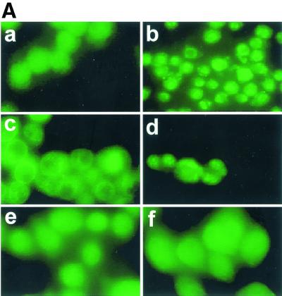
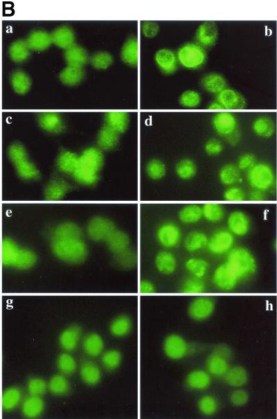
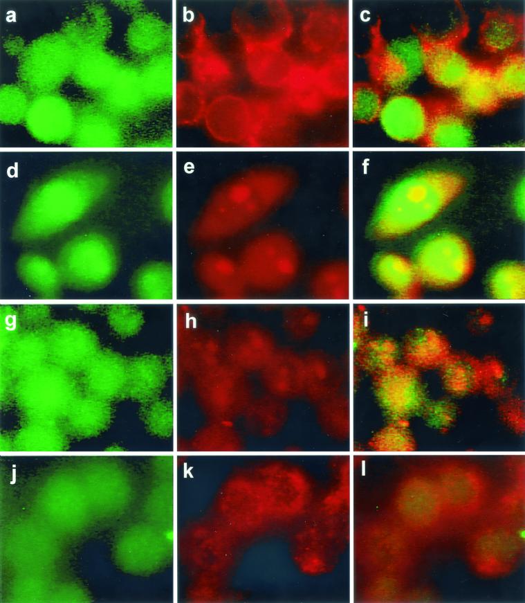
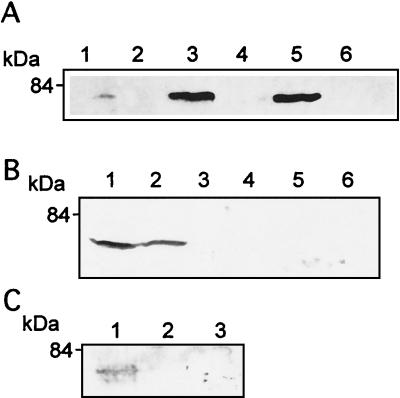
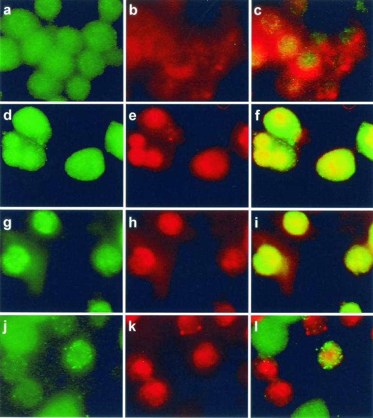
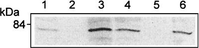
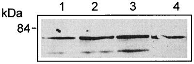
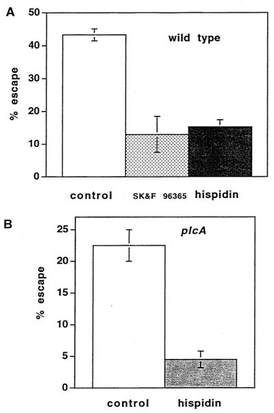
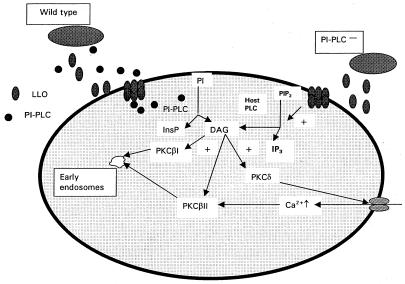
Listeriolysin O (LLO) and a phosphatidylinositol-specific phospholipase C (PI-PLC) are known virulence factors of Listeria monocytogenes in both tissue cultures and the murine model of infection. LLO is a member of a family of pore-forming cholesterol-dependent cytotoxins and is known to play an essential role in escape from the primary phagocytic vacuole of macrophages. PI-PLC plays an accessory role, in that PI-PLC mutants are partially defective in escape. We have shown that both of these molecules are essential for initiating rapid increases in the calcium level in the J774 murine macrophage cell line (S. J. Wadsworth and H. Goldfine, Infect. Immun. 67:1770-1778, 1999). Here we show that both LLO and PI-PLC are required for translocation of protein kinase C delta (PKC delta) to the periphery of J774 cells and for translocation of PKC beta II to early endosomes beginning within the first minute after addition of bacteria to the culture medium. Treatment with the calcium channel blocker SK&F 96365 inhibited translocation of PKC beta II but not PKC delta. Our findings lead us to propose a host signaling pathway requiring LLO and the formation of diacylglycerol by PI-PLC in which calcium-independent PKC delta is responsible for the initial calcium signal and the subsequent PKC beta II translocation. LLO-dependent translocation of PKC beta I to early endosomes also occurs between 1 and 4 min after infection, but this occurs in the absence of PI-PLC. All of these signals were observed in cells that had not internalized bacteria. Blocking PKC beta translocation with hispidin resulted in more rapid uptake of wild-type bacteria and greatly reduced escape from the primary phagocytic vacuoles of J774 cells.


Figures

FIG. 1.
FIG. 1.
Translocation of PKC δ after infection with L. monocytogenes or pretreatment with PMA. J774 cells were infected with different strains of L. monocytogenes or the same volume of PBS for 45 s and then processed for immunofluorescence analysis as described in the text. (A) Panel a, uninfected cells; panel b, uninfected cells treated with PMA (100 nM) for 10 min; panels c and d, cells infected with the wild type (PKC δ translocation is indicated by a bright green ring around the periphery of a cell [panel c] or by discrete bright round areas which appear to be clusters near the periphery of a cell [panel d]); panel e, cells infected with the LLO mutant strain; panel f, cells infected with the PI-PLC mutant strain. (B) Effects of inhibitors on translocation of PKC δ. PBS, SK&F 96365 (25 μM), hispidin (5 μM), or rottlerin (25 μM) was added to J774 cells 30 min prior to infection with the wild type. Uninfected cells to which PBS or inhibitor was added 30 min prior to infection are shown in the left panel of each row. Control or inhibitor-treated cells infected with the wild type for 45 s are shown in the right panel of each row. Panels a and b, control; panels c and d, cells pretreated with SK&F 96365; panels e and f, cells pretreated with hispidin; panels g and h, cells pretreated with rottlerin.
FIG. 1.
FIG. 1.
Translocation of PKC δ after infection with L. monocytogenes or pretreatment with PMA. J774 cells were infected with different strains of L. monocytogenes or the same volume of PBS for 45 s and then processed for immunofluorescence analysis as described in the text. (A) Panel a, uninfected cells; panel b, uninfected cells treated with PMA (100 nM) for 10 min; panels c and d, cells infected with the wild type (PKC δ translocation is indicated by a bright green ring around the periphery of a cell [panel c] or by discrete bright round areas which appear to be clusters near the periphery of a cell [panel d]); panel e, cells infected with the LLO mutant strain; panel f, cells infected with the PI-PLC mutant strain. (B) Effects of inhibitors on translocation of PKC δ. PBS, SK&F 96365 (25 μM), hispidin (5 μM), or rottlerin (25 μM) was added to J774 cells 30 min prior to infection with the wild type. Uninfected cells to which PBS or inhibitor was added 30 min prior to infection are shown in the left panel of each row. Control or inhibitor-treated cells infected with the wild type for 45 s are shown in the right panel of each row. Panels a and b, control; panels c and d, cells pretreated with SK&F 96365; panels e and f, cells pretreated with hispidin; panels g and h, cells pretreated with rottlerin.
FIG. 2.
FIG. 2.
Translocation of PKC β II to early endosomes of host J774 cells after infection with L. monocytogenes. PKC β II was labeled with FITC-conjugated secondary antibody as described in the text, and cells labeled for PKC β II are shown in the left panel of each row. Early endosomes labeled with rhodamine-conjugated transferrin are shown in the middle panel of each row. In uninfected cells these endosomes appear as discrete red areas near the periphery of a cell. In uninfected cells PKC β II is distributed throughout the cells and is also colocalized with early endosomes in ≤10% of the cells. The right panel of each row is the merged left and middle panels obtained by using the Phase 3 Imaging software. Upon infection with the wild type, PKC β II translocation to the early endosomes 45 s after addition of the bacteria appears as bright, discrete yellow areas in the same location as the early endosomes (f). PKC β II translocation did not occur after infection with mutant strains lacking LLO (g to i) or PI-PLC (j to l).
FIG. 3.
FIG. 3.
Isolation of early endosomes and Western blotting revealed translocation of PKC β II after infection. J774 cells were infected with the wild type or mutant strains of L. monocytogenes for 45 s, and purified endosome fractions were prepared. One hundred micrograms of protein of each sample (from two pooled endosome preparations) was used for SDS-polyacrylamide gel electrophoresis. (A) Infection with the wild type (lane 3) and the BR-PLC mutant strain (lane 5) resulted in increased PKC β II compared to the PKC β II in the uninfected cells (lane 1). Translocation of PKC β II was not observed in cells infected with the double phospholipase mutant (lane 2), the PI-PLC mutant (lane 4), or the LLO mutant (lane 6). (B) SK&F 96365 and hispidin inhibit translocation of PKC β II after infection with the wild type or the BR-PLC mutant strain. J774 cells were infected with the wild type (lane 1) or the BR-PLC mutant (lane 2), pretreated for 30 min with SK&F 91635 (25 μM) and infected with the wild type (lane 3) or the BR-PLC mutant (lane 4), or pretreated for 30 min with hispidin (5 μM) and infected with the wild type (lane 5) or the BR-PLC mutant (lane 6). (C) Rottlerin inhibits translocation of PKC β II after infection with the wild type. J774 cells were infected with the wild type (lane 1), pretreated for 30 min with hispidin (5 μM) and infected with wild type (lane 2), or pretreated for 30 min with rottlerin (25 μM) and infected with the wild type (lane 3).
FIG. 4.
FIG. 4.
PKC β I translocation occurred after infection with the wild type or the PI-PLC mutant strain but not after infection with the LLO mutant strain. PKC β I was labeled with FITC-conjugated secondary antibody as described in the text, and cells labeled for PKC β I are shown in the left panel of each row. Early endosomes were labeled with rhodamine-conjugated transferrin and appear in the middle panel of each row as discrete red areas. In uninfected cells PKC β I was located throughout the cytosol, could also appear as a discrete green area around the periphery of a cell, and could be colocalized with early endosomes. Cells were infected with the strains for 4 min. PKC β I translocation after infection with the wild-type strain (d to f) or the PI-PLC mutant strain (g to i) appears as discrete bright yellow areas corresponding to PKC β I colocalization with transferrin-labeled early endosomes. The images in the right panels were obtained as described in the legend to Fig. 2. Cells infected with the strain lacking LLO did not show translocation of PKC β I to early endosomes (j to l). Bright green objects seen especially in panels j and l are FITC-labeled bacteria.
FIG. 5.
FIG. 5.
Isolation of early endosomes and Western blotting revealed translocation of PKC β I after infection. J774 cells were infected with wild-type or mutant L. monocytogenes for 4 min, and purified endosome fractions were prepared. One hundred micrograms of protein of each sample (from two pooled endosome preparations) was used for SDS-polyacrylamide gel electrophoresis followed by Western blotting, as described in the text. PKC β I translocation was observed in cells infected with the wild type (lane 3) and the PI-PLC mutant (lane 6) compared to uninfected cells (lane 1). Translocation was not seen after infection with the LLO mutant (lane 2) or after 30 min of pretreatment with hispidin and infection with the wild type (lane 5). Cells pretreated with SK&F 96365 for 30 min showed translocation of PKC β I (lane 4).
FIG. 6.
FIG. 6.
Time course of translocation of PKC β I to early endosomes. Cells were infected with the wild type and harvested at intervals after infection. Early endosomes were isolated, and proteins were prepared for SDS-polyacrylamide gel electrophoresis and Western blotting as described in Materials and Methods. The proteins were also blotted with antibody to the transferrin receptor (upper bands). Infections were carried out for 1 min (lane 1), 3 min (lane 2), 4 min (lane 3), and 5 min (lane 4).
FIG. 7.
FIG. 7.
Effects of hispidin on association of L. monocytogenes with J774 cells and entry of L. monocytogenes into J774 cells. (A) Association of the wild type with untreated J774 cells (○) or cells pretreated with hispidin (5 μM) (⋄). (B) Association of the PI-PLC mutant with untreated J774 cells (□) or cells pretreated with hispidin (5 μM) (⋄). (C) Percentage of associated wild-type bacteria that were internalized in untreated J774 cells (○) or cells pretreated with hispidin (5 μM) (⋄). (D) Percentage of associated PI-PLC mutant bacteria that were internalized in untreated J774 cells (□) or cells pretreated for 30 min with hispidin (5 μM) (⋄). The total number of bacteria associated with the J774 cells was calculated as follows: total number of FITC-labeled bacteria (green)/total number of cells per image. The results are expressed as averages ± standard errors of the means for three sets of experiments in which a total of 200 to 250 J774 cells were counted. For association of the wild type in cells treated with hispidin P < 0.05 at 1, 5, and 10 min compared to the association for untreated cells. The percentage of internalized bacteria was calculated as described in the text. The results are expressed as averages ± standard errors of the means for three sets of experiments in which a total of 200 to 250 J774 cells were counted. For the percentage of internalized wild-type bacteria in cells treated with hispidin P < 0.05 compared to the percentage for untreated cells at 1, 5, and 10 min.
FIG. 8.
FIG. 8.
Hispidin pretreatment inhibited escape of the wild type and the PI-PLC mutant from the phagosome. Bacteria that escape from the phagosome and enter the cytosol become decorated with polymerized actin and can be stained with fluorescent phalloidin. After removal of uninternalized bacteria with gentamicin at 30 min, the percentage of cells that escaped at 1.5 h after infection was calculated as described in Materials and Methods. (A) Cells were either not pretreated (control) or pretreated with hispidin (5 μM) for 30 min and infected with the wild type. The data are the averages ± standard errors of the means for three experiments, and the difference is highly significant (P < 0.001). The data for cells pretreated with SK&F 96365 are taken from a previous study, in which the control gave an identical value (35). (B) Cells were pretreated with hispidin (5 μM) for 30 min and infected with the PI-PLC mutant. The data are averages ± standard errors of the means for three experiments, and the difference is highly significant (P < 0.01).
FIG. 9.
FIG. 9.
Suggested model for coupling of PKC activation and calcium signaling in host J774 cells after infection with L. monocytogenes. As described in the text, we suggest that PKC δ, calcium influx, and PKC β II activation are coupled and are initiated as a result of activity of two listerial proteins, LLO and PI-PLC. Activation of PKC β I did not require PI-PLC or elevated [Ca2+]i, and we suggest that it occurs as a result of the formation of DAG as a result of the action of host PLC on phosphatidylinositol 4,5-bisphosphate (PIP2) (12), yielding inositol 1,4,5-triphosphate (IP3).

Similar articles

Cited by

References

    1. Alvarez-Dominguez, C., A. M. Barbieri, W. Berón, A. Wandinger-Ness, and P. D. Stahl. 1996. Phagocytosed live Listeria monocytogenes influences rab5-regulated in vitro phagosome-endosome fusion. J. Biol. Chem. 271:13834-13843. - PubMed
    1. Bubert, A., Z. Sokolovic, S. K. Chun, L. Papatheodorou, A. Simm, and W. Goebel. 1999. Differential expression of Listeria monocytogenes virulence genes in mammalian host cells. Mol. Gen. Genet. 261:323-336. - PubMed
    1. Bucci, C., R. G. Parton, I. H. Mather, H. Stunnenberg, K. Simons, B. Hoflack, and M. Zerial. 1992. The small GTPase rab5 functions as a regulatory factor in the early endocytic pathway. Cell 70:715-728. - PubMed
    1. Camilli, A., H. Goldfine, and D. A. Portnoy. 1991. Listeria monocytogenes mutants lacking phosphatidylinositol-specific phospholipase C are avirulent. J. Exp. Med. 173:751-754. - PMC - PubMed
    1. Camilli, A., L. G. Tilney, and D. A. Portnoy. 1993. Dual roles of plcA in Listeria monocytogenes pathogenesis. Mol. Microbiol. 8:143-157. - PMC - PubMed

Publication types

MeSH terms

Substances