. 2002 Aug;70(8):4669-77.
doi: 10.1128/IAI.70.8.4669-4677.2002.

Escherichia coli shiga-like toxins induce apoptosis and cleavage of poly(ADP-ribose) polymerase via in vitro activation of caspases

Affiliations

Escherichia coli shiga-like toxins induce apoptosis and cleavage of poly(ADP-ribose) polymerase via in vitro activation of caspases

Joyce C Y Ching et al. Infect Immun. 2002 Aug.

Abstract

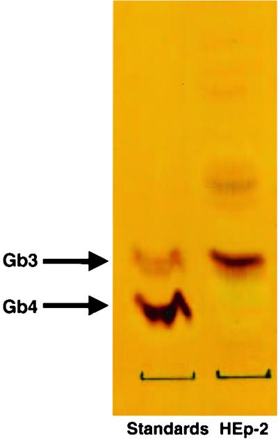
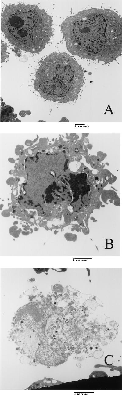
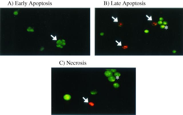
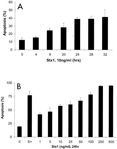
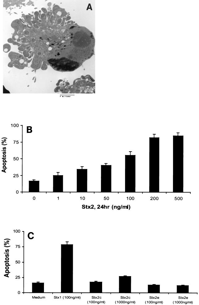
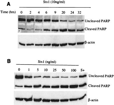
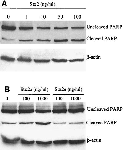
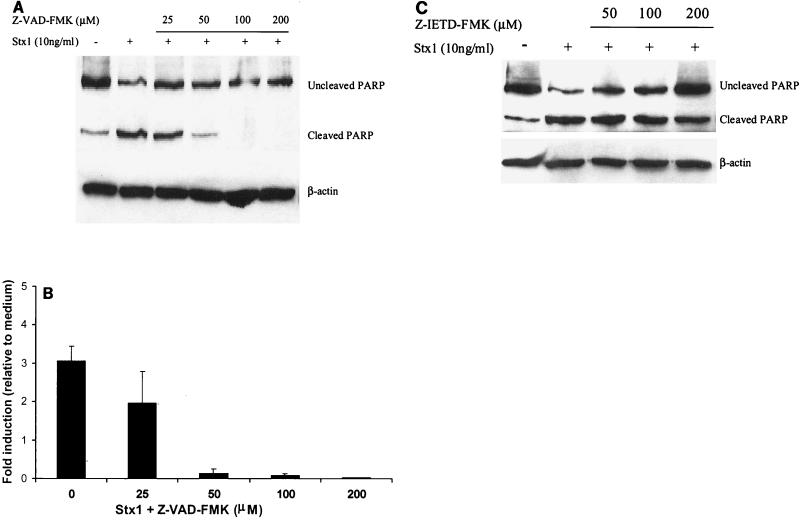
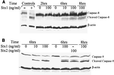
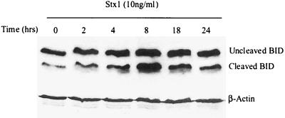
Shiga-like toxin-producing Escherichia coli causes hemorrhagic colitis and hemolytic-uremic syndrome in association with the production of Shiga-like toxins, which induce cell death via either necrosis or apoptosis. However, the abilities of different Shiga-like toxins to trigger apoptosis and the sequence of intracellular signaling events mediating the death of epithelial cells have not been completely defined. Fluorescent dye staining with acridine orange and ethidium bromide showed that Shiga-like toxin 1 (Stx1) induced apoptosis of HEp-2 cells in a dose- and time-dependent manner. Stx2 also induced apoptosis in a dose-dependent manner. Apoptosis induced by Stx1 (200 ng/ml) and apoptosis induced by Stx2 (200 ng/ml) were maximal following incubation with cells for 24 h (94.3% +/- 1.8% and 81.7% +/- 5.2% of the cells, respectively). Toxin-treated cells showed characteristic features of apoptosis, including membrane blebbing, DNA fragmentation, chromatin condensation, cell shrinkage, and the formation of apoptotic bodies, as assessed by transmission electron microscopy. Stx2c induced apoptosis weakly even at a high dose (1,000 ng/ml for 24 h; 26.7% +/- 1.3% of the cells), whereas Stx2e did not induce apoptosis of HEp-2 cells. Thin-layer chromatography confirmed that HEp-2 cells express the Stx1-Stx2-Stx2c receptor, globotriaosylceramide (Gb3), but not the Stx2e receptor, globotetraosylceramide (Gb4). Western blot analysis of poly(ADP-ribose) polymerase (PARP), a DNA repair enzyme, demonstrated that incubation with Stx1 and Stx2 induced cleavage, whereas incubation with Stx2e did not result in cleavage of PARP. A pan-caspase inhibitor (Z-VAD-FMK) and a caspase-8-specific inhibitor (Z-IETD-FMK) eliminated, in a dose-dependent fashion, the cleavage of PARP induced by Shiga-like toxins. Caspase-8 activation was confirmed by detection of cleavage of this enzyme by immunoblotting. Cleavage of caspase-9 and the proapoptotic member of the Bcl-2 family BID was also induced by Stx1, as determined by immunoblot analyses. We conclude that different Shiga-like toxins induce different degrees of apoptosis that correlates with toxin binding to the glycolipid receptor Gb3 and that caspases play an integral role in the signal transduction cascade leading to toxin-mediated programmed cell death.


Figures

FIG. 1.
FIG. 1.
Gb3, and not Gb4, is present in HEp-2 cells, as determined by thin-layer chromatography of extracted lipids from HEp-2 cells.
FIG. 2.
FIG. 2.
Stx1 induces apoptosis of HEp-2 cells, as assessed by transmission electron microscopy. (A) HEp-2 cells grown in medium alone, displaying normal morphology. (B) HEp-2 cells treated with Stx1 (100 ng/ml) for 24 h, displaying membrane blebbing, chromatin condensation, DNA fragmentation, and the formation of apoptotic bodies, consistent with apoptosis. (C) Necrotic HEp-2 cell after Shiga-like toxin treatment, showing loss of membrane integrity, cell swelling, and leakage of cytoplasmic contents. The micrographs are electron micrographs that are representative of at least three separate experiments. Bars = 2 μm.
FIG. 3.
FIG. 3.
Stx1-induced apoptosis, as assessed by acridine orange and ethidium bromide fluorescent dye staining. (A) Early apoptotic HEp-2 cell (arrow), showing the presence of green patches of fragmented and condensed chromatin. (B) Late apoptotic cells fluoresce orange (arrows), whereas viable cells are uniformly green (asterisk). As the plasma membrane loses its integrity, ethidium bromide enters the cell and intercalates fragmented DNA, staining the cell red. (C) Necrotic cell (arrow) that has lost its selective permeability, allowing ethidium bromide to intercalate DNA and produce a uniform red color. Viable cells are uniformly green (asterisk). Original approximate magnifications, ×400.
FIG. 4.
FIG. 4.
Stx1 induces apoptosis in HEp-2 cells in a time- and dose-dependent manner. (A) Apoptosis was quantified by fluorescent dye staining of HEp-2 cells treated with Stx1 (10 ng/ml) for various times (n = 3 for each time point; P < 0.005, as determined by ANOVA). (B) HEp-2 cells treated with various concentrations of Stx1 for 24 h. Apoptosis was quantified by fluorescent dye staining. Staurosporine (1 μM) (bar S+), a known inducer of apoptosis (4), was used as a positive control. Untreated cells (bar 0) served as the negative control (n ≥ 6; P < 0.001, as determined by ANOVA).
FIG. 5.
FIG. 5.
Induction of apoptosis by Stx2 and Stx2c in HEp-2 cells. (A) Transmission electron micrograph showing a HEp-2 cell treated with Stx2 (100 ng/ml) for 24 h undergoing apoptosis, as shown by the characteristic morphological features of apoptosis. Bar = 2 μm. (B) Fluorescence microscopy showed that incubation with Stx2 for 24 h induced apoptosis in a dose-dependent manner (n ≥ 6; P < 0.001, as determined by ANOVA). (C) The Stx2 variant Stx2c (1,000 ng/ml) (n = 5; P < 0.001, as determined by ANOVA), but not Stx2e, induced apoptosis of HEp-2 cells.
FIG. 6.
FIG. 6.
Stx1 induces PARP cleavage in a time- and dose-dependent manner, as determined by Western blot analysis of HEp-2 whole-cell protein lysates probed with PARP antibody. (A) Immunoblot of HEp-2 lysates following incubation with Stx1 (10 ng/ml), demonstrating PARP cleavage in a time-dependent fashion. Membranes were probed for β-actin to determine protein loading in each lane. (B) Immunoblot showing Stx1 (10 ng/ml)-induced PARP cleavage in HEp-2 cells in a dose-dependent manner. Staurosporine (1,000 nM) (lane S+) was used as a positive control. Membranes were probed for β-actin to determine protein loading in each lane.
FIG. 7.
FIG. 7.
Stx2 induces PARP cleavage in a dose-dependent manner, whereas Stx2e does not. (A) Immunoblot showing PARP cleavage in HEp-2 cells following 24 h of treatment with various concentrations of Stx2. Membranes were probed for β-actin to determine protein loading in each lane. (B) Stx2c (1,000 ng/ml) induces PARP cleavage, whereas Stx2e does not induce cleavage of PARP. Membranes were probed for β-actin to determine protein loading in each lane.
FIG. 8.
FIG. 8.
Pan-caspase and specific caspase-8 inhibitors eliminate Stx1-induced PARP cleavage. (A) Pretreatment of HEp-2 cells with the pan-caspase inhibitor Z-VAD-FMK for 2 h at 37°C prior to Stx1 (10 ng/ml) treatment for 24 h eliminated PARP cleavage induced by the toxin. Membranes were probed for β-actin to determine protein loading in each lane. (B) Densitometry of Western blot of Stx1-mediated PARP cleavage inhibited by Z-VAD-FMK. The results of three individual experiments are shown (P < 0.01, as determined by ANOVA). (C) Pretreatment of HEp-2 cells with the caspase-8 inhibitor Z-IETD-FMK 2 h prior to toxin treatment for 24 h resulted in decreased PARP cleavage induced by Stx1.
FIG. 9.
FIG. 9.
Caspase-8 and caspase-9 are activated by Stx1. (A) Western blot analysis of HEp-2 whole-cell protein lysates probed with caspase-8 antibody. Lanes − and + contained control HeLa cells that were not treated and treated with cycloheximide-tumor necrosis factor (Cell Signaling Technologies), respectively. Incubation of HEp-2 cells with Stx1 (100 ng/ml) for 2 h did not induce cleavage of caspase-8. However, treatment of HEp-2 cells with Stx1 (10 and 100 ng/ml) for 6 and 8 h resulted in cleavage of caspase-8, demonstrating that caspase-8 cleavage occurred in a time-dependent fashion. Membranes were probed for β-actin to determine protein loading in each lane. (B) Whole-cell protein lysates probed with caspase-9 antibody. Treatment of HEp-2 cells with Stx1 (10 and 100 ng/ml) or Stx2 (100 ng/ml) for 6 h induced cleavage of caspase-9. Membranes were probed for β-actin to determine protein loading in each lane.
FIG. 10.
FIG. 10.
Immunoblot demonstrating that Stx1 induces cleavage of BID in HEp-2 cells. Membranes were stripped and reprobed for β-actin to assess protein loading in each lane.

Similar articles

Cited by

References

    1. Abreu, M. T., A. A. Palladino, E. T. Arnold, R. S. Kwon, and J. A. McRoberts. 2000. Modulation of barrier function during Fas-mediated apoptosis in human intestinal epithelial cells. Gastroenterology 119:1524-1536. - PubMed
    1. Agbodaze, D. 1999. Verocytotoxins (Shiga-like toxins) produced by Escherichia coli: a minireview of their classification, clinical presentations and management of a heterogeneous family of cytotoxins. Comp. Immunol. Microbiol. Infect. Dis. 22:221-230. - PubMed
    1. Arab, S., M. Murakami, P. Dirks, B. Boyd, S. L. Hubbard, C. A. Lingwood, and J. T. Rutka. 1998. Verotoxins inhibit the growth of and induce apoptosis in human astrocytoma cells. J. Neurooncol. 40:137-150. - PubMed
    1. Belmokhtar, C. A., J. Hillion, and E. Segal-Bendirdjian. 2001. Staurosporine induces apoptosis through both caspase-dependent and caspase-independent mechanisms. Oncogene 20:3354-3362. - PubMed
    1. Braun, J. S., R. Novak, K. H. Herzog, S. M. Bodner, J. L. Cleveland, and E. I. Tuomanen. 1999. Neuroprotection by a caspase inhibitor in acute bacterial meningitis. Nat. Med. 5:298-302. - PubMed

Publication types

MeSH terms

Substances

LinkOut - more resources