. 2002 Jul 15;196(2):237-46.
doi: 10.1084/jem.20020590.

CD4(+)CD25(+) regulatory T cells can mediate suppressor function in the absence of transforming growth factor beta1 production and responsiveness

Affiliations

CD4(+)CD25(+) regulatory T cells can mediate suppressor function in the absence of transforming growth factor beta1 production and responsiveness

Ciriaco A Piccirillo et al. J Exp Med. .

Abstract

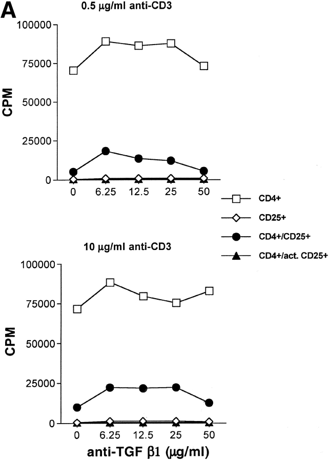
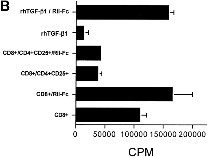
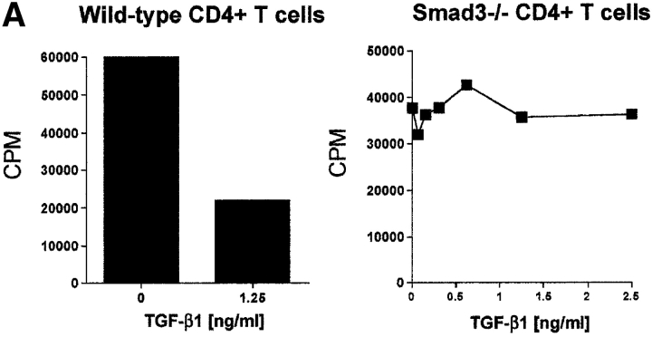
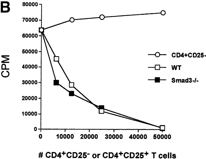
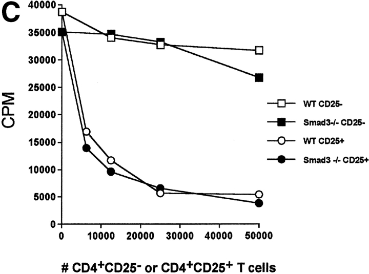
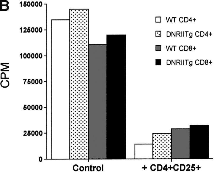
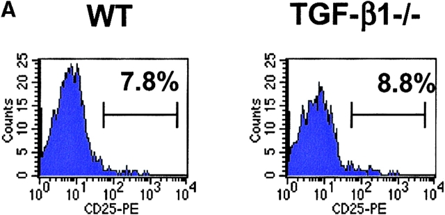
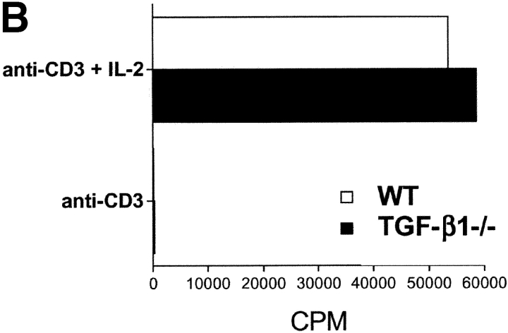
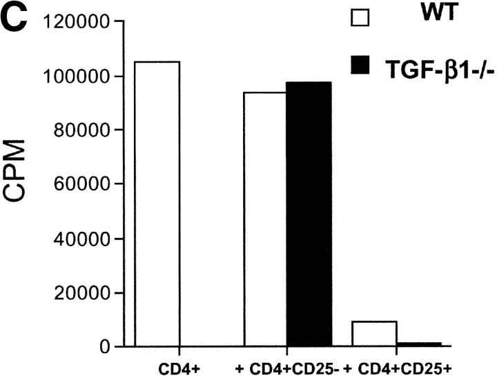
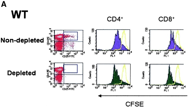
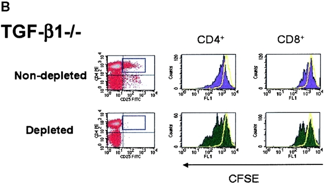
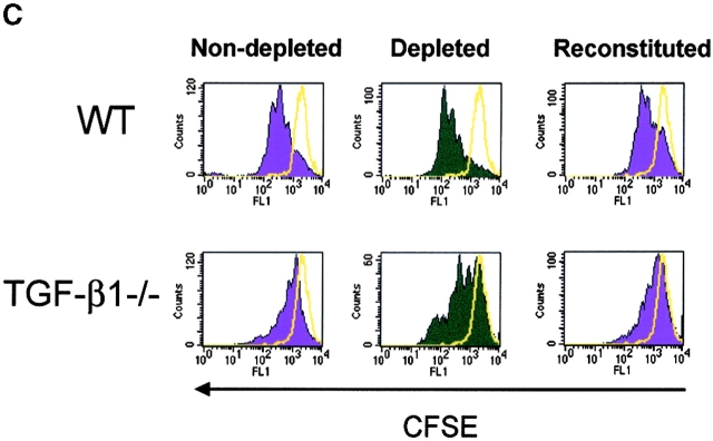
CD4(+)CD25(+) regulatory T cells inhibit organ-specific autoimmune diseases induced by CD4(+)CD25(-) T cells and are potent suppressors of T cell activation in vitro. Their mechanism of suppression remains unknown, but most in vitro studies suggest that it is cell contact-dependent and cytokine independent. The role of TGF-beta1 in CD4(+)CD25(+) suppressor function remains unclear. While most studies have failed to reverse suppression with anti-transforming growth factor (TGF)-beta1 in vitro, one recent study has reported that CD4(+)CD25(+) T cells express cell surface TGF-beta1 and that suppression can be completely abrogated by high concentrations of anti-TGF-beta suggesting that cell-associated TGF-beta1 was the primary effector of CD4(+)CD25(+)-mediated suppression. Here, we have reevaluated the role of TGF-beta1 in CD4(+)CD25(+)-mediated suppression. Neutralization of TGF-beta1 with either monoclonal antibody (mAb) or soluble TGF-betaRII-Fc did not reverse in vitro suppression mediated by resting or activated CD4(+)CD25(+) T cells. Responder T cells from Smad3(-/-) or dominant-negative TGF-beta type RII transgenic (DNRIITg) mice, that are both unresponsive to TGF-beta1-induced growth arrest, were as susceptible to CD4(+)CD25(+)-mediated suppression as T cells from wild-type mice. Furthermore, CD4(+)CD25(+) T cells from neonatal TGF-beta1(-/-) mice were as suppressive as CD4(+)CD25(+) from TGF-beta1(+/+) mice. Collectively, these results demonstrate that CD4(+)CD25(+) suppressor function can occur independently of TGF-beta1.


Figures

Figure 1.
Figure 1.
TGF-β1 blockade does not reverse CD4+CD25+-mediated suppression of T cell proliferation. (A) WT CD4+ T cells (5 × 10⁴) were stimulated with irradiated T cell–depleted spleen cells (2 × 10⁵) either alone or with freshly isolated or activated (•) CD4+CD25+ T cells (1:2 suppressor/responder ratio) in the presence of titrated doses of anti–TGF-β1. Results from a representative experiment of three are shown. (B) OT-I Tg CD8+ T cells were stimulated with H-2Kb/OVA257–264 soluble tetramer (0.1 μg/ml) either alone or with activated CD4+CD25+ T cells (1:2 suppressor/responder ratio) in the presence or absence of soluble TGF-βRII-Fc (10 μg/ml). In some instances, rhTGF-β1 was added as a control (5 ng/ml).
Figure 1.
Figure 1.
TGF-β1 blockade does not reverse CD4+CD25+-mediated suppression of T cell proliferation. (A) WT CD4+ T cells (5 × 10⁴) were stimulated with irradiated T cell–depleted spleen cells (2 × 10⁵) either alone or with freshly isolated or activated (•) CD4+CD25+ T cells (1:2 suppressor/responder ratio) in the presence of titrated doses of anti–TGF-β1. Results from a representative experiment of three are shown. (B) OT-I Tg CD8+ T cells were stimulated with H-2Kb/OVA257–264 soluble tetramer (0.1 μg/ml) either alone or with activated CD4+CD25+ T cells (1:2 suppressor/responder ratio) in the presence or absence of soluble TGF-βRII-Fc (10 μg/ml). In some instances, rhTGF-β1 was added as a control (5 ng/ml).
Figure 2.
Figure 2.
CD4+ T cells from TGF-β1–insensitive Smad3−/− mice are susceptible to suppression mediated by CD4+CD25+ T cells. (A) WT or Smad3−/− CD4+ T cells (5 × 10⁴) were stimulated with anti-CD3 (0.5 μg/ml) and irradiated, T cell–depleted spleen cells (2 × 10⁵) in the presence or absence of exogenous rhTGF-β1. (B) WT or Smad3−/− CD4+ T cells (5 × 10⁴) were stimulated as described in A in the presence of titrated numbers of either activated CD4+CD25+ or CD4+CD25 T cells. (C) Smad3-dependent signaling in CD4+CD25+ T cells is not required for suppressor effector function. WT CD4+ T cells were stimulated as described in A and in the presence of either freshly isolated CD4+CD25+ or CD4+CD25 T cells from WT or Smad3−/− mice. The final purity of sorted suppressors was >98%.
Figure 2.
Figure 2.
CD4+ T cells from TGF-β1–insensitive Smad3−/− mice are susceptible to suppression mediated by CD4+CD25+ T cells. (A) WT or Smad3−/− CD4+ T cells (5 × 10⁴) were stimulated with anti-CD3 (0.5 μg/ml) and irradiated, T cell–depleted spleen cells (2 × 10⁵) in the presence or absence of exogenous rhTGF-β1. (B) WT or Smad3−/− CD4+ T cells (5 × 10⁴) were stimulated as described in A in the presence of titrated numbers of either activated CD4+CD25+ or CD4+CD25 T cells. (C) Smad3-dependent signaling in CD4+CD25+ T cells is not required for suppressor effector function. WT CD4+ T cells were stimulated as described in A and in the presence of either freshly isolated CD4+CD25+ or CD4+CD25 T cells from WT or Smad3−/− mice. The final purity of sorted suppressors was >98%.
Figure 3.
Figure 3.
CD4+CD25+ T cells suppress T cells from DNRIITg mice. (A) T cells from WT or DNRIITg mice (5 × 10⁴ cells) were stimulated with anti-CD3 (0.5 μg/ml) and irradiated, T cell–depleted spleen cells (2 × 10⁵) in the presence or absence of exogenous rhTGF-β1. (B) CD4+ or CD8+ T cells (5 × 10⁴) from WT or DNRIITg mice were stimulated as described in A in the presence or absence of either activated CD4+CD25+ or CD4+CD25 T cells.
Figure 3.
Figure 3.
CD4+CD25+ T cells suppress T cells from DNRIITg mice. (A) T cells from WT or DNRIITg mice (5 × 10⁴ cells) were stimulated with anti-CD3 (0.5 μg/ml) and irradiated, T cell–depleted spleen cells (2 × 10⁵) in the presence or absence of exogenous rhTGF-β1. (B) CD4+ or CD8+ T cells (5 × 10⁴) from WT or DNRIITg mice were stimulated as described in A in the presence or absence of either activated CD4+CD25+ or CD4+CD25 T cells.
Figure 4.
Figure 4.
CD4+CD25+ regulatory T cell activity is operative in TGF-β1−/− mice. (A) The frequency of CD4+CD25+ T cells is comparable in WT and TGF-β1−/− neonatal mice. (B) WT or TGF-β1−/− CD4+ CD25+ or CD4+CD25 T cells (5 × 10⁴) were stimulated with anti-CD3 (0.5 μg/ml) and irradiated, T cell–depleted spleen cells (2 × 10⁵) in the presence or absence of exogenous IL-2 (5 ng/ml). (C) WT CD4+ T cells were stimulated as described in B in the presence of either freshly isolated CD4+CD25+ or CD4+ CD25 T cells from WT or TGF-β1−/− neonatal mice.
Figure 4.
Figure 4.
CD4+CD25+ regulatory T cell activity is operative in TGF-β1−/− mice. (A) The frequency of CD4+CD25+ T cells is comparable in WT and TGF-β1−/− neonatal mice. (B) WT or TGF-β1−/− CD4+ CD25+ or CD4+CD25 T cells (5 × 10⁴) were stimulated with anti-CD3 (0.5 μg/ml) and irradiated, T cell–depleted spleen cells (2 × 10⁵) in the presence or absence of exogenous IL-2 (5 ng/ml). (C) WT CD4+ T cells were stimulated as described in B in the presence of either freshly isolated CD4+CD25+ or CD4+ CD25 T cells from WT or TGF-β1−/− neonatal mice.
Figure 5.
Figure 5.
In vitro depletion of CD4+CD25+ regulatory T cell activity in TGF-β1−/− mice enhances T cell proliferation. CD25-depleted or nondepleted splenocytes/lymph node cells from either WT (A) or TGF-β1−/− (B) neonatal mice were labeled with CFSE and stimulated with anti-CD3 (0.5 μg/ml) and irradiated, T cell–depleted spleen cells (2 × 10⁵) for a period of 48 h. Cells were then analyzed by flow cytometry for CFSE division status relative to unstimulated T cells (solid yellow line). All histograms were gated on CD4+ or CD8+ T cells. (C) CD25-depleted WT or TGF-β1−/− cells were reconstituted with the respective nondepleted cell preparation, stimulated with soluble anti-CD3, and then examined the frequency of dividing cells based on CFSE dilution by flow cytometry. Cells were gated on CD4+ T cells.
Figure 5.
Figure 5.
In vitro depletion of CD4+CD25+ regulatory T cell activity in TGF-β1−/− mice enhances T cell proliferation. CD25-depleted or nondepleted splenocytes/lymph node cells from either WT (A) or TGF-β1−/− (B) neonatal mice were labeled with CFSE and stimulated with anti-CD3 (0.5 μg/ml) and irradiated, T cell–depleted spleen cells (2 × 10⁵) for a period of 48 h. Cells were then analyzed by flow cytometry for CFSE division status relative to unstimulated T cells (solid yellow line). All histograms were gated on CD4+ or CD8+ T cells. (C) CD25-depleted WT or TGF-β1−/− cells were reconstituted with the respective nondepleted cell preparation, stimulated with soluble anti-CD3, and then examined the frequency of dividing cells based on CFSE dilution by flow cytometry. Cells were gated on CD4+ T cells.
Figure 6.
Figure 6.
Anti–TGF-β does not reverse suppression of AIG mediated by CD4+CD25+ T cells. Nu/nu mice (6–8 wk) were injected with splenocytes depleted of CD25+ cells (20 × 106 cells), as described in the Materials and Methods. Simultaneously, some nu/nu mice were coinjected with CD4+CD25+ T cells (2 × 10⁶ cells). Anti–TGF-β (2 mg) was injected on days –1 and 1 of cell transfer and weekly thereafter for the duration of the experiment. Stomachs were harvested 6 wk after transfer and H&E stains of formalin-fixed, paraffin-embedded sections were analyzed for gastric pathology.

References

    1. Bach, J.F. 1995. Organ-specific autoimmunity. Immunol. Today. 16:353–355. - PubMed
    1. King, C., and N. Sarvetnick. 1997. Organ-specific autoimmunity. Curr. Opin. Immunol. 9:863–871. - PubMed
    1. Moller, G. 1996. Dominant immunological tolerance. Immunol. Rev. 149:1–17.
    1. Shevach, E.M. 2000. Regulatory T cells in autoimmmunity. Annu. Rev. Immunol. 18:423–449. - PubMed
    1. Sakaguchi, S. 2000. Regulatory T cells: key controllers of immunologic self-tolerance. Cell. 101:455–458. - PubMed

MeSH terms

Substances