. 2002 Jul 15;196(2):247-53.
doi: 10.1084/jem.20020642.

Human CD4(+)CD25(+) regulatory, contact-dependent T cells induce interleukin 10-producing, contact-independent type 1-like regulatory T cells [corrected]

Affiliations

Human CD4(+)CD25(+) regulatory, contact-dependent T cells induce interleukin 10-producing, contact-independent type 1-like regulatory T cells [corrected]

Detlef Dieckmann et al. J Exp Med. .

Erratum in

  • J Exp Med 2002 Aug 19;196(4):559
  • J Exp Med 2002 Sep 16;196(6):867

Abstract

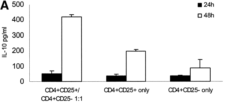
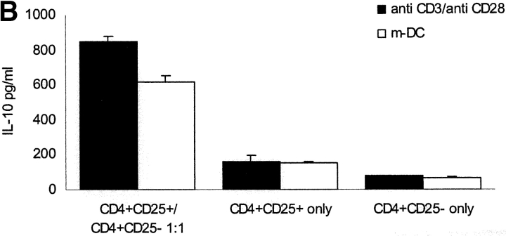
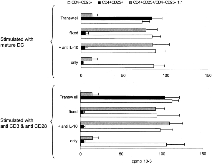
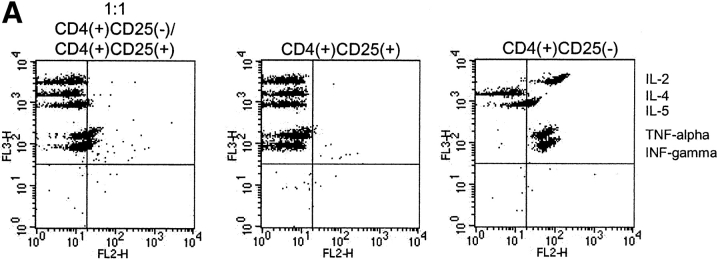
It has been recently demonstrated that regulatory CD4(+)CD25(+) CD45RO(+) T cells are present in the peripheral blood of healthy adults and exert regulatory function similar to their rodent counterparts. It remains difficult to understand how the small fraction of these T cells that regulate via direct cell-to-cell contact and not via secretion of immunosuppressive cytokines could mediate strong immune suppression. Here we show that human CD4(+)CD25(+) T cells induce long-lasting anergy and production of interleukin (IL)-10 in CD4(+)CD25(-) T cells. These anergized CD4(+)CD25(-) T cells then suppress proliferation of syngenic CD4(+) T cells via IL-10 but independent of direct cell contact, similar to the so-called type 1 regulatory T (Tr1) cells. This 'catalytic' function of CD4(+)CD25(+) T cells to induce Tr1-like cells helps to explain their central role for the maintenance of immune homeostasis.


Figures

Figure 1.
Figure 1.
Coculture of CD4+CD25+ and CD4+CD25 T cells results in high level IL-10 production. CD4+CD25+ and CD4+CD25 T cells were MACS® sorted from PBMCs of healthy individuals. These cells were either cultured alone or at a 1:1 ratio and activated with platebound anti-CD3 and soluble anti-CD28 (10 μg/ml, respectively). (A) After various time points supernatants were analyzed for cytokine production by ELISA. IL-10 production peaked 48 h after onset of culture and was markedly higher in the coculture of CD4+CD25+ and CD4+CD25 T cells than in the cultures of each of the cell types alone. A representative out of five independent standardized experiments is shown. No elevated levels of INF-α or TGF-β could be measured (data not shown). (B) The different T cell populations were also activated with mature allogeneic DCs (DC/T cell ratio 1:20) compared with anti-CD3 and anti-CD28 (10 μg/ml, respectively). Cytokines were measured 48 h after onset of culture. Results were similar in five independent experiments. (C) For the last 6 h of activation with anti-CD3 and anti-CD28 2 μM monensin was added to the cultures. Staining of CD4 surface expression was performed. Cells were washed, fixed, permeabilized, and stained for intracellular IL-10 using PE-conjugated specific Abs. One of five independent experiments is shown.
Figure 1.
Figure 1.
Coculture of CD4+CD25+ and CD4+CD25 T cells results in high level IL-10 production. CD4+CD25+ and CD4+CD25 T cells were MACS® sorted from PBMCs of healthy individuals. These cells were either cultured alone or at a 1:1 ratio and activated with platebound anti-CD3 and soluble anti-CD28 (10 μg/ml, respectively). (A) After various time points supernatants were analyzed for cytokine production by ELISA. IL-10 production peaked 48 h after onset of culture and was markedly higher in the coculture of CD4+CD25+ and CD4+CD25 T cells than in the cultures of each of the cell types alone. A representative out of five independent standardized experiments is shown. No elevated levels of INF-α or TGF-β could be measured (data not shown). (B) The different T cell populations were also activated with mature allogeneic DCs (DC/T cell ratio 1:20) compared with anti-CD3 and anti-CD28 (10 μg/ml, respectively). Cytokines were measured 48 h after onset of culture. Results were similar in five independent experiments. (C) For the last 6 h of activation with anti-CD3 and anti-CD28 2 μM monensin was added to the cultures. Staining of CD4 surface expression was performed. Cells were washed, fixed, permeabilized, and stained for intracellular IL-10 using PE-conjugated specific Abs. One of five independent experiments is shown.
Figure 2.
Figure 2.
Activated fixed CD4+CD25+ T cells show similar regulatory potential as viable CD4+CD25+ T cells and can induce IL-10 production in CD4+CD25 T cells. (A) CD4+ T cell subpopulations were sorted by MACS® CD4+CD25+ T cells were divided into three fractions. One part was activated with platebound anti-CD3 (10 μg/ml) and soluble anti-CD28 (10 μg/ml) overnight and fixed the next day with paraformaldehyde 2% (activated-fixed). The third part was fixed with paraformaldehyde without activation (resting-fixed) and the second part was left untreated (viable). Each fraction was mixed with syngeneic CD4+CD25 T cells at a 1:1 ratio (105 T cells per 96 well) and stimulated with platebound anti-CD3 (10 μg/ml) and soluble-anti-CD28 (10 μg/ml). Proliferation was determined by 3[H]Tdr incorporation after 5 d. Results are representative of five independent experiments, shown as mean cpm of triplicate cultures. Similar results were observed when T cells were stimulated with mature allogeneic DCs (DC/T cell ratio of 1:20; data not shown) (B) CD4+CD25+ and CD4+CD25 T cells were either cultured alone or CD4+CD25 T cells were mixed at a 1:1 ratio with activated-fixed, resting-fixed or viable CD4+CD25+ T cells. T cells were stimulated with mature allogeneic DCs at the same ratio as in A. In a parallel transwell approach CD4+CD25+ T cells were stimulated with allogeneic DCs (DC/T ratio 1:20) in a transwell chamber, and CD4+CD25 T cells were placed in the well together with allogeneic DCs again at a DC/T ratio of 1:20. IL-10 production was measured by ELISA 48 h after onset of culture. Results were similar in five independent experiments.
Figure 2.
Figure 2.
Activated fixed CD4+CD25+ T cells show similar regulatory potential as viable CD4+CD25+ T cells and can induce IL-10 production in CD4+CD25 T cells. (A) CD4+ T cell subpopulations were sorted by MACS® CD4+CD25+ T cells were divided into three fractions. One part was activated with platebound anti-CD3 (10 μg/ml) and soluble anti-CD28 (10 μg/ml) overnight and fixed the next day with paraformaldehyde 2% (activated-fixed). The third part was fixed with paraformaldehyde without activation (resting-fixed) and the second part was left untreated (viable). Each fraction was mixed with syngeneic CD4+CD25 T cells at a 1:1 ratio (105 T cells per 96 well) and stimulated with platebound anti-CD3 (10 μg/ml) and soluble-anti-CD28 (10 μg/ml). Proliferation was determined by 3[H]Tdr incorporation after 5 d. Results are representative of five independent experiments, shown as mean cpm of triplicate cultures. Similar results were observed when T cells were stimulated with mature allogeneic DCs (DC/T cell ratio of 1:20; data not shown) (B) CD4+CD25+ and CD4+CD25 T cells were either cultured alone or CD4+CD25 T cells were mixed at a 1:1 ratio with activated-fixed, resting-fixed or viable CD4+CD25+ T cells. T cells were stimulated with mature allogeneic DCs at the same ratio as in A. In a parallel transwell approach CD4+CD25+ T cells were stimulated with allogeneic DCs (DC/T ratio 1:20) in a transwell chamber, and CD4+CD25 T cells were placed in the well together with allogeneic DCs again at a DC/T ratio of 1:20. IL-10 production was measured by ELISA 48 h after onset of culture. Results were similar in five independent experiments.
Figure 3.
Figure 3.
CD4+CD25 T cells anergized by CD4+CD25+ T cells suppress proliferation of CD4+ T cells in a IL-10–dependent manner. MACS® sorted CD4+CD25+ and CD4+CD25 T cells were either cultured alone or mixed at a 1:1 ratio (2 × 10⁶ T cells per 24 well) and stimulated with mature allogeneic DCs (DC/T cell ratio 1:20) or immobilized anti-CD3/soluble anti-CD28. After 48 h of culture cells were harvested and one fraction of each population was fixed with paraformaldehyde for 1 h. Viable and fixed cells were cocultured with syngeneic resting CD4+CD25 T cells at a 1:1 ratio (105 T cells per 96 well) and stimulated as before with immobilized anti-CD3/soluble anti-CD28 (bottom panel) or mature allogeneic DCs (top panel) in the presence or absence of 10 μg/ml anti–IL-10 Abs. In a parallel transwell approach the three different T cell populations were placed in a transwell chamber and resting CD4+CD25 T cells were stimulated with DCs (DC/T cell ratio 1:20; top panel) or platebound anti-CD3/soluble anti-CD28 (bottom panel) in the well. Proliferation after 5 d was determined by 3[HT]Tdr incorporation. One out of four independent experiments is shown.
Figure 4.
Figure 4.
(A) Anergized CD4+CD25 T cells predominantly secrete IL-10. CD4+CD25+ and CD4+CD25 T cells were isolated as described and stimulated alone or at a 1:1 ratio with anti-CD3/anti-CD28. 48 h after stimulation supernatant was harvested and analyzed by a cytometric bead array for IL-2, IL-4, IL-5, TNF-α, and INF-γ. Results were similar in five independent experiments. (B) Before mixing CD4+CD25 and CD4+CD25+ T cells at a 1:1 ratio, CD4+CD25+ T cells were labeled with 0.5 μM CFSE for 15 min. Cells were then mixed and stimulated with immobilized anti-CD3/soluble anti-CD28. After 48 h cells were harvested and sorted on a FACSVantage™. The positive and the negative fraction were then cocultured with syngeneic resting CD4+CD25 T cells (105 T cells per 96 well). Proliferation was measured after 5 d by 3[HT]Tdr incorporation. One out of five independent experiments is shown.
Figure 4.
Figure 4.
(A) Anergized CD4+CD25 T cells predominantly secrete IL-10. CD4+CD25+ and CD4+CD25 T cells were isolated as described and stimulated alone or at a 1:1 ratio with anti-CD3/anti-CD28. 48 h after stimulation supernatant was harvested and analyzed by a cytometric bead array for IL-2, IL-4, IL-5, TNF-α, and INF-γ. Results were similar in five independent experiments. (B) Before mixing CD4+CD25 and CD4+CD25+ T cells at a 1:1 ratio, CD4+CD25+ T cells were labeled with 0.5 μM CFSE for 15 min. Cells were then mixed and stimulated with immobilized anti-CD3/soluble anti-CD28. After 48 h cells were harvested and sorted on a FACSVantage™. The positive and the negative fraction were then cocultured with syngeneic resting CD4+CD25 T cells (105 T cells per 96 well). Proliferation was measured after 5 d by 3[HT]Tdr incorporation. One out of five independent experiments is shown.

References

    1. Asano, M., M. Toda, N. Sakaguchi, and S. Sakaguchi. 1996. Autoimmune disease as a consequence of developmental abnormality of a T cell subpopulation. J. Exp. Med. 184:387–396. - PMC - PubMed
    1. Takahashi, T., Y. Kuniyasu, M. Toda, N. Sakaguchi, M. Itoh, M. Iwata, J. Shimizu, and S. Sakaguchi. 1998. Immunologic self-tolerance maintained by CD25+CD4+ naturally anergic and suppressive T cells: induction of autoimmune disease by breaking their anergic/suppressive state. Int. Immunol. 10:1969–1980. - PubMed
    1. Thornton, A.M., and E.M. Shevach. 1998. CD4+CD25+ immunoregulatory T cells suppress polyclonal T cell activation in vitro by inhibiting interleukin 2 production. J. Exp. Med. 188:287–296. - PMC - PubMed
    1. Jonuleit, H., E. Schmitt, M. Stassen, A. Tuettenberg, J. Knop, and A.H. Enk. 2001. Identification and functional characterization of human CD4+CD25+ T cells with regulatory properties isolated from peripheral blood. J. Exp. Med. 193:1285–1294. - PMC - PubMed
    1. Levings, M.K., R. Sangregorio, and M.G. Roncarolo. 2001. Human CD25+CD4+ T regulatory cells suppress naive and memory T cell proliferation and can be expanded in vitro without loss of function. J. Exp. Med. 193:1295–1302. - PMC - PubMed

Publication types

Substances