. 2002 Jul 22;158(2):209-14.
doi: 10.1083/jcb.200203103. Epub 2002 Jul 15.

A phosphatidylinositol (4,5)-bisphosphate binding site within mu2-adaptin regulates clathrin-mediated endocytosis

Affiliations

A phosphatidylinositol (4,5)-bisphosphate binding site within mu2-adaptin regulates clathrin-mediated endocytosis

Gundula Rohde et al. J Cell Biol. .

Abstract

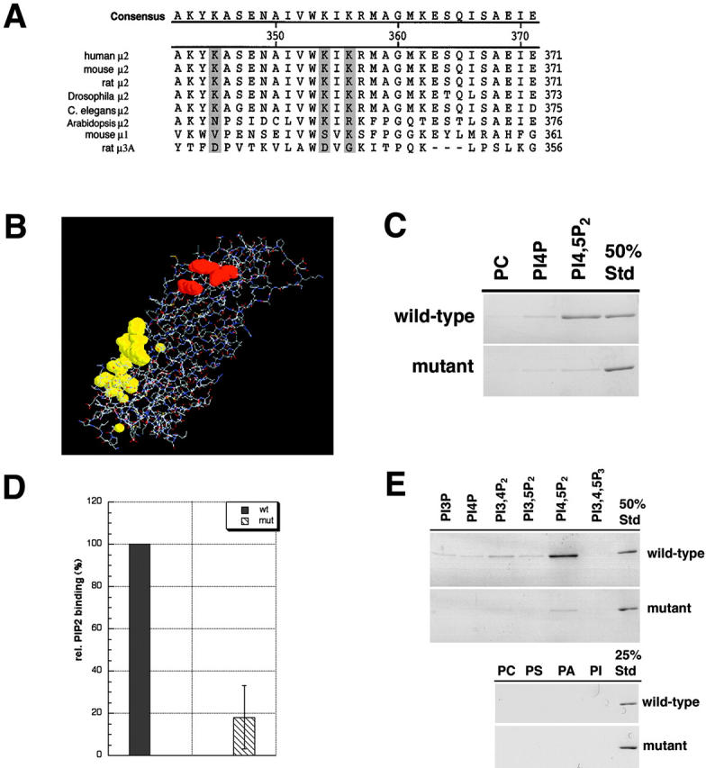
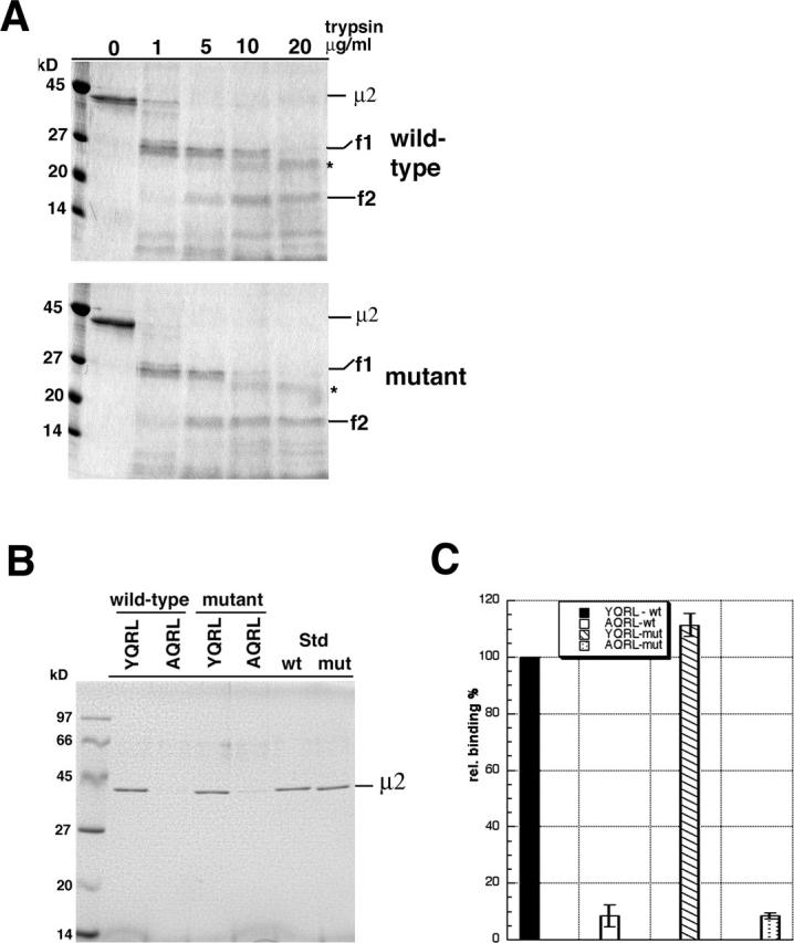
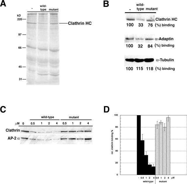
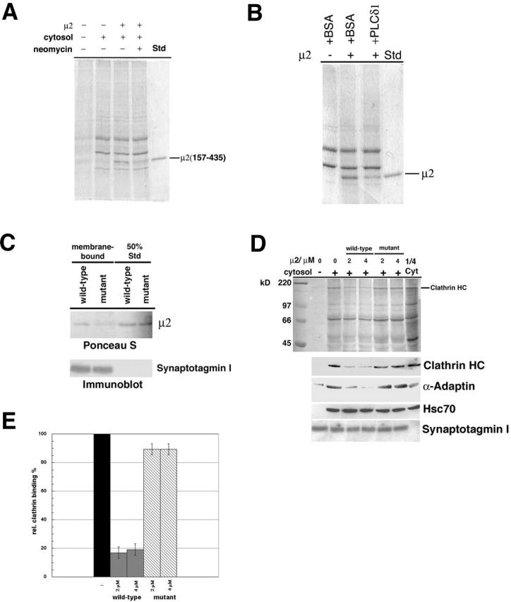
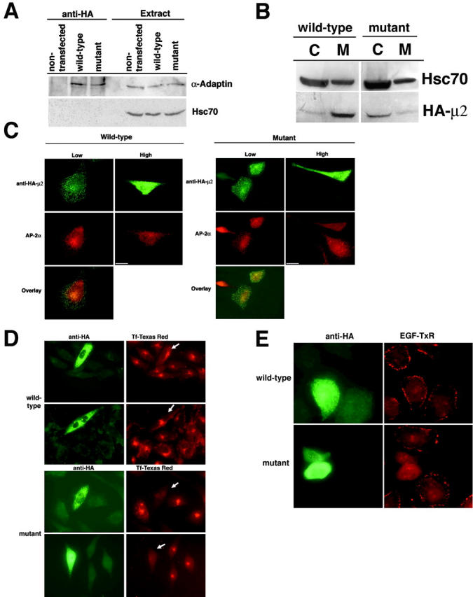
The clathrin adaptor complex AP-2 serves to coordinate clathrin-coated pit assembly with the sorting of transmembrane cargo proteins at the plasmalemma. How precisely AP-2 assembly and cargo protein recognition at sites of endocytosis are regulated has remained unclear, but recent evidence implicates phosphoinositides, in particular phosphatidylinositol (4,5)-bisphosphate (PI[4,5]P2), in these processes. Here we have identified and functionally characterized a conserved binding site for PI(4,5)P2 within mu2-adaptin, the medium chain of the clathrin adaptor complex AP-2. Mutant mu2 lacking a cluster of conserved lysine residues fails to bind PI(4,5)P2 and to compete the recruitment of native clathrin/AP-2 to PI(4,5)P2-containing liposomes or to presynaptic membranes. Moreover, we show that expression of mutant mu2 inhibits receptor-mediated endocytosis in living cells. We suggest that PI(4,5)P2 binding to mu2-adaptin regulates clathrin-mediated endocytosis and thereby may contribute to structurally linking cargo recognition to coat formation.


Figures

Figure 1.
Figure 1.
Phosphoinositide binding of μ2-adaptin. (A) Sequence alignment of μ-adaptins from various species. The conserved lysine residues within the putative phosphoinositide binding site are shaded. (B) Structural model of μ2 (aa 157–435) complexed with a tyrosine-based endocytosis signal (yellow) (modified from Owen and Evans, 1998). The three conserved lysine residues (red) form a basic patch at the surface of subdomain B. (C) Purified wild-type or mutant μ2 (2 μg) were incubated with liposomes containing 10% PC, PI(4)P, or PI(4,5)P2. After reisolation of the liposomes, samples were analyzed by SDS-PAGE and staining with Coomassie blue. 50% Std, 50% of the protein sample added to the assay. (D) Quantification of the binding of wild-type or mutant μ2 to PI(4,5) P2-containing liposomes from three independent experiments and plotted as mean ± SE. (E) Binding of μ2 to liposomes containing 10% of the indicated lipid. PS, phosphatidylserine; PA, phosphatidic acid. The experiment was performed as described in A.
Figure 2.
Figure 2.
Effects of the KKK–EEE mutation on the interaction of μ2 with tyrosine-based endocytosis signals and synaptotagmin. (A) μ2 (157–435; 2 μg) was incubated with the indicated concentrations of trypsin for 15 min at RT. Samples were analyzed by 12% SDS-PAGE and staining with Coomassie blue. The asterisk denotes trypsin. (B) Mutant and wild-type μ2 (157–435; 2 μg) were incubated for 1 h at 4°C with peptides bearing the tyrosine-based endocytosis motif of TGN38 (YQRL) or its AQRL mutant immobilized on beads. Beads were reisolated, washed, and analyzed by SDS-PAGE and staining with Coomassie blue. Std, 50% of the protein added to the assay. (C) Quantification of the results shown in B. Data from three independent experiments were analyzed and plotted as mean ± SE. Binding of wild-type μ2 to the YQRL peptide was set as 100%.
Figure 3.
Figure 3.
Recruitment of clathrin/AP-2 to PI(4,5)P2-containing liposomes. (A and B) PI(4,5)P2-containing liposomes were incubated with cytosol, ATP, GTPγS, and 4 μM wild-type or mutant μ2 (157–435). Liposomes were reisolated, washed, and analyzed by SDS-PAGE and staining with Ponceau S (A) or immunoblotting (B). Bands were visualized with 125I-protein A and quantified by phosphoimage analysis. (C) Dose dependence of μ2-mediated inhibition of clathrin/AP-2 recruitment to liposomes. The experiment was done as described in A, using the indicated concentrations of wild-type or mutant μ2. (D) Quantification of clathrin recruitment onto liposomes in the presence of different concentrations of μ2. Data are plotted as mean (±SE) from several experiments. The amount of clathrin recruited to liposomes in the absence of added μ2 was taken as 100%.
Figure 4.
Figure 4.
Recruitment of clathrin/AP-2 and μ2 to synaptic LP2 membranes. (A) Membrane association of μ2 is inhibited by neomycin. Carbonate-washed LP2 membranes (10 μg) were incubated with cytosol (0.4 mg/ml), ATP, GTPγS, and μ2 (157–435; 1.5 μg) in the presence or absence of 2 mM neomycin. LP2 membranes were reisolated, washed, and analyzed by Western blotting and staining with Ponceau S. Std, 50% of the μ2 added to the assay. (B) Membrane association of μ2 is inhibited by phospholipase Cδ1. Recruitment of μ2 was assayed as described in A in the presence or absence of 5 μg purified phospholipase Cδ1 or BSA. (C) Membrane recruitment of μ2 requires an intact PI(4,5)P2-binding site. Recruitment of wild-type or mutant μ2 was analyzed as in A, except that the samples were analyzed by staining with Ponceau S to detect bound μ2 and immunoblotting for synaptotagmin I as a membrane marker. (D) Clathrin/AP-2 recruitment to LP2 membranes can be competed by wild-type but not KKK–EEE mutant μ2. LP2 membranes (20 μg) were incubated with cytosol, ATP, GTPγS, and 2 or 4 μM of wild-type or mutant μ2 (157–435). Membranes were reisolated, washed, and analyzed by staining with Ponceau S (top) or immunoblotting (bottom) for clathrin heavy chain (HC), α-adaptin, hsc70, and synaptotagmin I. 1/4 cyt, 25% of the cytosol used in the experiment. (E) Quantification of clathrin recruitment as shown in D. The amount of clathrin recruited to LP2 in the absence of μ2 was taken as 100%. Data are plotted as mean (±SE) from three independent experiments.
Figure 5.
Figure 5.
Expression of KKK–EEE mutant μ2 inhibits clathrin-mediated endocytosis in CHO cells. (A) CHO cells (10 × 106) transiently transfected with HA-tagged wild-type or mutant μ2 were lysed and subjected to immunoprecipitations with monoclonal antibodies against the HA tag. Samples were analyzed by SDS-PAGE and immunoblotting for α-adaptin and hsc70. Extract, 10% of the total extracted proteins used for the experiment. (B) Transfected CHO cells (as in A) were fractionated into membrane (M) and cytosol (C). Samples were analyzed by SDS-PAGE and immunoblotting for HA-tagged μ2 or hsc70. (C) HA-tagged wild-type or mutant μ2 were transiently expressed in CHO cells. 48 h after transfection, cells were methanol fixed and immunostained with antibodies against α-adaptin or HA. Bar, 10 μm. (D) HA-tagged wild-type or mutant μ2 was transiently expressed in CHO cells (see C) and analyzed for their ability to internalize Texas red–labeled transferrin (2.5 μg/ml; 10 min at 37°C) by immunofluorescence microscopy. Transfected cells are indicated by an arrow. The results are representative of three independent transfection experiments in which 85% of the cells expressing elevated levels of mutant μ2 displayed strongly reduced transferrin uptake. (E) HA-tagged wild-type or mutant μ2 were transiently expressed in HeLa cells and analyzed for the ability to internalize Texas red–labeled EGF (2 μg/ml; 3 min at 37°C) by immunofluorescence microscopy.

References

    1. Aguilar, R.C., H. Ohno, K.W. Roche, and J.S. Bonifacino. 1997. Functional domain mapping of the clathrin-associated adaptor medium chains mu1 and mu2. J. Biol. Chem. 272:27160–27166. - PubMed
    1. Arneson, L.S., J. Kunz, R.A. Anderson, and L.M. Traub. 1999. Coupled inositide phosphorylation and phospholipase D activation initiates clathrin-coat assembly on lysosomes. J. Biol. Chem. 274:17794–17805. - PubMed
    1. Brodin, L., P. Low, and O. Shupliakov. 2000. Sequential steps in clathrin-mediated synaptic vesicle endocytosis. Curr. Opin. Neurobiol. 10:312–320. - PubMed
    1. Bromann, P.A., E.E. Boetticher, and J.W. Lomasney. 1997. A single amino acid substitution in the pleckstrin homology domain of phospholipase C delta1 enhances the rate of substrate hydrolysis. J. Biol. Chem. 272:16240–16246. - PubMed
    1. Cremona, O., G. Di Paolo, M.R. Wenk, A. Luthi, W.T. Kim, K. Takei, L. Daniell, Y. Nemoto, S.B. Shears, R.A. Flavell, et al. 1999. Essential role of phosphoinositide metabolism in synaptic vesicle recycling. Cell. 99:179–188. - PubMed

Publication types

MeSH terms

Substances