. 2002 Jul 22;158(2):227-33.
doi: 10.1083/jcb.200204026. Epub 2002 Jul 15.

Autotaxin has lysophospholipase D activity leading to tumor cell growth and motility by lysophosphatidic acid production

Affiliations

Autotaxin has lysophospholipase D activity leading to tumor cell growth and motility by lysophosphatidic acid production

Makiko Umezu-Goto et al. J Cell Biol. .

Abstract

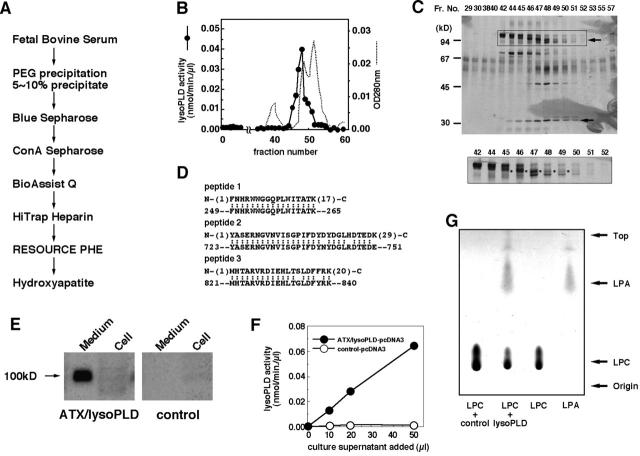
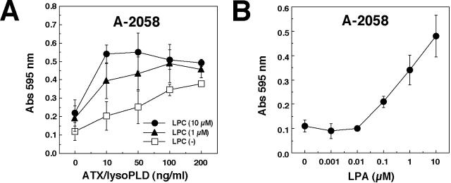
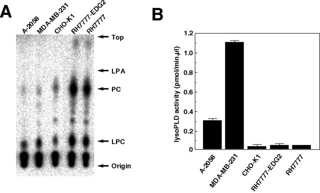
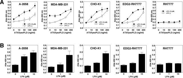
Autotaxin (ATX) is a tumor cell motility-stimulating factor, originally isolated from melanoma cell supernatants. ATX had been proposed to mediate its effects through 5'-nucleotide pyrophosphatase and phosphodiesterase activities. However, the ATX substrate mediating the increase in cellular motility remains to be identified. Here, we demonstrated that lysophospholipase D (lysoPLD) purified from fetal bovine serum, which catalyzes the production of the bioactive phospholipid mediator, lysophosphatidic acid (LPA), from lysophosphatidylcholine (LPC), is identical to ATX. The Km value of ATX for LPC was 25-fold lower than that for the synthetic nucleoside substrate, p-nitrophenyl-tri-monophosphate. LPA mediates multiple biological functions including cytoskeletal reorganization, chemotaxis, and cell growth through activation of specific G protein-coupled receptors. Recombinant ATX, particularly in the presence of LPC, dramatically increased chemotaxis and proliferation of multiple different cell lines. Moreover, we demonstrate that several cancer cell lines release significant amounts of LPC, a substrate for ATX, into the culture medium. The demonstration that ATX and lysoPLD are identical suggests that autocrine or paracrine production of LPA contributes to tumor cell motility, survival, and proliferation. It also provides potential novel targets for therapy of pathophysiological states including cancer.


Figures

Figure 1.
Figure 1.
Purification of lysoPLD and identification as ATX. (A) Strategy used for purification of lysoPLD. (B) Elution profile of lysoPLD activity on hydroxylapatite column chromatography (CHT-II). The flow-through fractions of the RESOURCE® PHE hydrophobic column were applied to a CHT-II column, and the absorbed proteins were eluted with a linearly increasing gradient of KH2PO4 (0–0.4 M). 1.5 ml-fractions were collected, examined for lysoPLD activity (closed circle), and protein concentration was monitored by absorbance at 280 nm (dashed line). (C) The active fractions from the CHT-II column were subjected to SDS-PAGE and the proteins were detected by silver staining. Arrows indicate the protein bands that co-migrate with lysoPLD activity. The lower band (∼30 kD) was a degradation product of the 100-kD band as judged by mass spectrometry and sequence analyses (unpublished data). In the bottom panel, an enlarged picture of the active fractions (fractions 42–52) is shown. The 100-kD band is indicated by an asterisk. (D) Alignment of peptide sequences obtained from amino acid sequence analysis of bovine lysoPLD with the amino acid sequence of rat autotaxin. Numbers in the second lines correspond to the amino acid numbers of rat ATX. Identical amino acids were indicated by colons. (E) Expression of ATX/lysoPLD recombinant protein in CHO-K1 cells. Culture supernatant or cells from myc epitope–tagged ATX/lysoPLD-pcDNA3 or control pcDNA3 vector transfected CHO-K1 were subjected to Western blot analysis using anti-myc mAb (9E11). ATX/lysoPLD protein was recovered almost exclusively from culture supernatant of ATX/lysoPLD-pcDNA3– transfected cells. (F) Recombinant ATX shows lysoPLD activity as assessed by choline release. The culture supernatant of ATX/lysoPLD-pcDNA3 (closed circle) or control pcDNA3 vector–transfected CHO-K1 (open circle) was subjected to a lysoPLD assay. (G) Formation LPA by lysoPLD. Phospholipids in the lysoPLD reaction (F) were analyzed by TLC after the lipids in the reaction mixture were extracted by organic solvents. LPC or LPA is included to indicate migration on the TLC plate.
Figure 2.
Figure 2.
ATX/lysoPLD-induced chemotaxis is enhanced by lysophosphatidylcholine in A-2058 melanoma cells. (A) Lysophosphatidylcholine (LPC) enhances chemotaxis of A-2058 cells induced by recombinant ATX/lysoPLD. ATX/lysoPLD-induced chemotaxis was assessed in the presence or absence of LPC (1-oleoyl, 1 μM; closed triangle) or 10 μM (closed circle; mean ± SEM, n = 4). ATX/lysoPLD was produced by baculovirus (see Materials and methods). (B) Dose dependency of LPA (1-oleoyl)-induced chemotaxis of A-2058 and CHO-K1 cells (mean ± SEM, n = 4; see Materials and methods).
Figure 4.
Figure 4.
Secretion of LPC and expression of lysoPLD activity by a variety of cell lineages. (A) Monolayers of cell lines were preincubated with [32P]orthophosphoric acid for 12 h. Lipids in the culture medium were extracted with chloroform and methanol, and then subjected to TLC. Radioactivity of each lipid spot was detected and visualized using an image analyzer. Identification of each lipid spot was confirmed by using standard lipids both in one- and two-dimensional TLC (unpublished data). (B) Cancer cells secrete lysoPLD. Cells were cultured for 48 h in serum-free medium, and the lysoPLD activity of the culture medium was determined (see Materials and methods). The following cells were used: A-2058, MDA-MB-231, CHO-K1, RH7777-EDG2, and parental RH7777.
Figure 3.
Figure 3.
ATX/lysoPLD stimulates cell proliferation. (A) ATX/lysoPLD stimulates cell growth of cancer cell lines and LPA receptor–expressing rat hepatoma cells. Cells were starved for 48 h. Cell proliferation induced by the addition of recombinant ATX/lysoPLD both in the presence or absence of 10 μM LPC (1-oleoyl) was evaluated by MTT hydrolysis (mean ± SEM, n = 3). The following cells were used: A-2058 (melanoma, human), MDA-MB-231 (breast cancer, human), CHO-K1 (ovary-derived fibroblast, hamster), RH7777-EDG2, and parental RH7777 (hepatoma, rat). (B) Dose dependency of LPA (1-oleoyl)-induced cell growth. LPA-induced cell growth was assessed by MTT hydrolysis as in A (mean ± SEM, n = 3).

Comment in

References

    1. An, S., T. Bleu, O.G. Hallmark, and E.J. Goetzl. 1998. Characterization of a novel subtype of human G protein-coupled receptor for lysophosphatidic acid. J. Biol. Chem. 273:7906–7910. - PubMed
    1. Bandoh, K., J. Aoki, H. Hosono, S. Kobayashi, T. Kobayashi, M.K. Murakami, M. Tsujimoto, H. Arai, and K. Inoue. 1999. Molecular cloning and characterization of a novel human G-protein-coupled receptor, EDG7, for lysophosphatidic acid. J. Biol. Chem. 274:27776–27785. - PubMed
    1. Bligh, E.C., and W.F. Dyer. 1959. A rapid method for total lipid extraction and purification. Can. J. Biochem. Physiol. 37:911–917. - PubMed
    1. Bollen, M., R. Gijsbers, H. Ceulemans, W. Stalmans, and C. Stefan. 2000. Nucleotide pyrophosphatases/phosphodiesterases on the move. Crit. Rev. Biochem. Mol. Biol. 35:393–432. - PubMed
    1. Clair, T., H.Y. Lee, L.A. Liotta, and M.L. Stracke. 1997. Autotaxin is an exoenzyme possessing 5′-nucleotide phosphodiesterase/ATP pyrophosphatase and ATPase activities. J. Biol. Chem. 272:996–1001. - PubMed

Publication types

MeSH terms