. 2002 Jul 22;158(2):215-20.
doi: 10.1083/jcb.200204066. Epub 2002 Jul 15.

Mossy fiber Zn2+ spillover modulates heterosynaptic N-methyl-D-aspartate receptor activity in hippocampal CA3 circuits

Affiliations

Mossy fiber Zn2+ spillover modulates heterosynaptic N-methyl-D-aspartate receptor activity in hippocampal CA3 circuits

Sayaka Ueno et al. J Cell Biol. .

Abstract

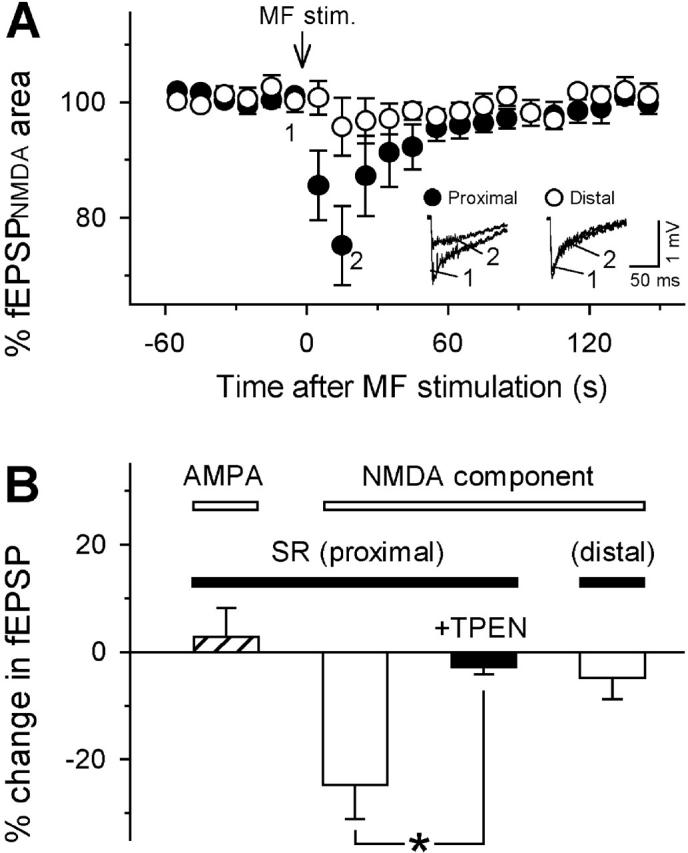
Although Zn2+ is contained in large amounts in the synaptic terminals of hippocampal mossy fibers (MFs), its physiological role in synaptic transmission is poorly understood. By using the newly developed high-sensitivity Zn2+ indicator ZnAF-2, the spatiotemporal dynamics of Zn2+ was monitored in rat hippocampal slices. When high-frequency stimulation was delivered to the MFs, the concentration of extracellular Zn2+ was immediately elevated in the stratum lucidum, followed by a mild increase in the stratum radiatum adjacent to the stratum lucidum, but not in the distal area of stratum radiatum. The Zn2+ increase was insensitive to a non-N-methyl-d-aspartate (NMDA) receptor antagonist but was efficiently attenuated by tetrodotoxin or Ca2+-free medium, suggesting that Zn2+ is released by MF synaptic terminals in an activity-dependent manner, and thereafter diffuses extracellularly into the neighboring stratum radiatum. Electrophysiological analyses revealed that NMDA receptor-mediated synaptic responses in CA3 proximal stratum radiatum were inhibited in the immediate aftermath of MF activation and that this inhibition was no longer observed in the presence of a Zn2+-chelating agent. Thus, Zn2+ serves as a spatiotemporal mediator in imprinting the history of MF activity in contiguous hippocampal networks. We predict herein a novel form of metaplasticity, i.e., an experience-dependent non-Hebbian modulation of synaptic plasticity.


Figures

Figure 1.
Figure 1.
ZnAF-2 labels endogenous Zn2 + in rat hippocampal slices. (A) Confocal image of a hippocampal slice loaded with a membrane-permeable, diacetylated form of ZnAF-2. (B) Confocal image of the boxed region in A taken at higher magnification. The fluorescent signal of ZnAF-2 was detected in subgranular zone, dentate hilus (DH), stratum lucidum (SL) and a small portion of stratum oriens (SO) but not seen in stratum radiatum (SR) or stratum pyramidale (SP), which corresponds to intracellular Zn2+ localization. ZnAF-2 images of the CA3 area in a hippocampal slice were obtained immediately before (C) and 15 min (D) after bath application of 25 μM TPEN. ZnAF-2 signal was eliminated by Zn2+ chelation by TPEN.
Figure 2.
Figure 2.
Extracellular Zn2 + release and diffusion after MF activation. (A) An image of the dentato-CA3 area of a hippocampal slice perfused with ZnAF-2. Confocal ZnAF-2 signal is shown as a green-colorized scale, superimposed on a transmitted beam image. Bipolar electrodes (*) were placed in stratum granulosum (SG) to stimulate the MFs. The dotted line marks the transect of illumination during line-scan imaging. (B) Line-scan image of ZnAF-2 taken at the points indicated in A. The temporal resolution was 1 s per line. “Hotter” colors correspond to increased [Zn2+]o on an arbitrary pseudo-color scale. (C) Data extracted from the image in B, along the time axis. Each point in time is the average %ΔF/F value across the spatial axis of the region separated by the horizontal dotted lines in B, i.e., the stratum radiatum far from stratum lucidum (SR distal, brown), the stratum radiatum near stratum lucidum (SR proximal, green), stratum lucidum (SL, red) and stratum pyramidale (SP, blue). The MFs were tetanized at 100 Hz for 2 s (MF stim.) at the time indicated by the vertical dotted line. MF stimulation elicited an immediate increase in [Zn2+]o in stratum lucidum, and a slow increase in stratum pyramidale and proximal stratum radiatum but no apparent change in distal stratum radiatum. D, The area between the white dotted lines (a 0.02 mm2 square) was strongly illuminated in the presence of 200 μM Zn2+ for photobleaching and imaged 30 s before and 0, 25 and 60 s after the illumination. Fluorescence recovery owing to a diffusion of Zn2+-ZnAF-2 complex was not found within at least 60 s. E, Summary data of %ΔF/F 15 s after MF stimulation in stratum lucidum (red) and proximal (green) and distal (brown) stratum radiatum in the absence (Control) or presence of 2 μM tetrodotoxin (TTX) or 20 μM CNQX, or in Ca2+-free medium. *P < 0.01 versus Control; Student's t test. Data are means ± SEM of 5–13 slices.
Figure 3.
Figure 3.
Frequency-dependent dynamics of synaptically released Zn2 + . (A–C) Time course of %ΔF/F of ZnAF-2 in stratum lucidum (SL, red), proximal stratum radiatum (SR proximal, green) and distal stratum radiatum (SR distal, brown) after MF stimulation of 200 pulses at 100 Hz (A), 200 pulses 5 Hz (B) and 60 pulses at 1 Hz (C). The stimulation was applied from time 0. While the 100 Hz stimulation evoked a rapid, large increase in [Zn2+]o, the lower frequencies of stimulation caused a slow, moderate increase. (D) Basal ΔF/F (no MF stimulation) was stable. Data represent means ± SEM of 5–13 slices.
Figure 4.
Figure 4.
Zn2 + -mediated, heterosynaptic inhibition of NMDA receptors in stratum radiatum after MF activation. (A) fEPSPNMDA in associational/commissural fiber-CA3 pyramidal cell synapses were extracellularly recorded from the stratum radiatum proximal to (closed circles) or far from (open circles) stratum lucidum in Mg2+-free solution containing 20 μM CNQX. When MF stimulation (100 Hz for 2 s) was applied at time from −2 to 0 (MF stim.), the NMDA component was temporarily depressed. Representative traces of fEPSPNMDA at times −5 and 15 are shown in the insets. (B) Summary data for the effect of MF stimulation on AMPA and NMDA responses in proximal and distal stratum radiatum (SR). The ordinate indicates an average change in fEPSPs 15 s after MF stimulation (100 Hz for 2 s). The fEPSPNMDA depression was blocked by 25 μM TPEN. Neither fEPSPAMPA in proximal stratum radiatum nor fEPSPNMDA in distal |stratum radiatum was unaffected by MF activation. *P < 0.01; Student's t test. Data are means ± SEM of 5–7 slices.

References

    1. Assaf, S.Y., and S.H. Chung. 1984. Release of endogenous Zn2+ from brain tissue during activity. Nature. 308:734–736. - PubMed
    1. Bliss, T.V., and G.L. Collingridge. 1993. A synaptic model of memory: long-term potentiation in the hippocampus. Nature. 361:31–39. - PubMed
    1. Brewer, G.J., J.C. Aster, C.A. Knutsen, and W.C. Kruckeberg. 1979. Zinc inhibition of calmodulin: a proposed molecular mechanism of zinc action on cellular functions. Am. J. Hematol. 7:53–60. - PubMed
    1. Canzoniero, L.M., D.M. Turetsky, and D.W. Choi. 1999. Measurement of intracellular free zinc concentrations accompanying zinc-induced neuronal death. J. Neurosci. 19:RC31. - PMC - PubMed
    1. Choi, D.W., and J.Y. Koh. 1998. Zinc and brain injury. Annu. Rev. Neurosci. 21:347–375. - PubMed

Publication types