. 2002 Jul;14(7):1541-55.
doi: 10.1105/tpc.001156.

Nucleocytoplasmic partitioning of the plant photoreceptors phytochrome A, B, C, D, and E is regulated differentially by light and exhibits a diurnal rhythm

Affiliations

Nucleocytoplasmic partitioning of the plant photoreceptors phytochrome A, B, C, D, and E is regulated differentially by light and exhibits a diurnal rhythm

Stefan Kircher et al. Plant Cell. 2002 Jul.

Abstract

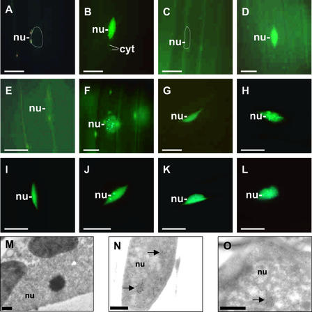
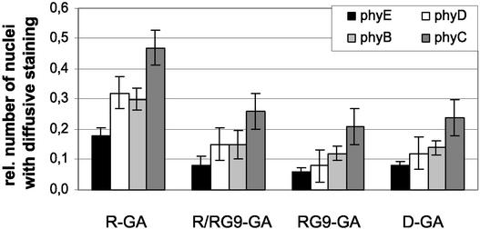
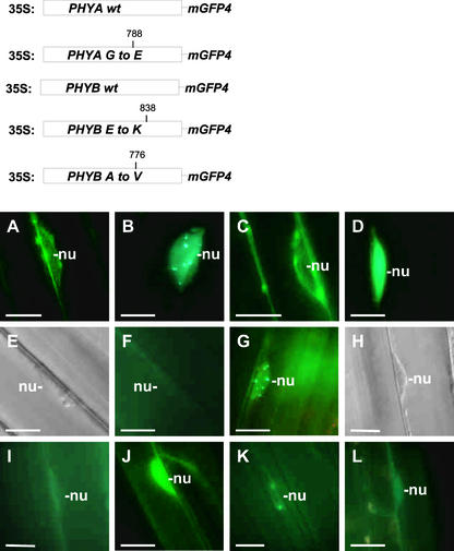
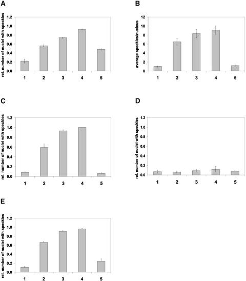
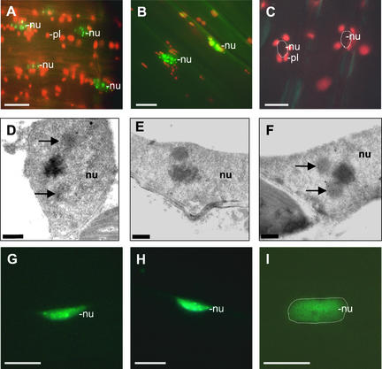
The phytochrome family of plant photoreceptors has a central role in the adaptation of plant development to changes in ambient light conditions. The individual phytochrome species regulate different or partly overlapping physiological responses. We generated transgenic Arabidopsis plants expressing phytochrome A to E:green fluorescent protein (GFP) fusion proteins to assess the biological role of intracellular compartmentation of these photoreceptors in light-regulated signaling. We show that all phytochrome:GFP fusion proteins were imported into the nuclei. Translocation of these photoreceptors into the nuclei was regulated differentially by light. Light-induced accumulation of phytochrome species in the nuclei resulted in the formation of speckles. The appearance of these nuclear structures exhibited distinctly different kinetics, wavelengths, and fluence dependence and was regulated by a diurnal rhythm. Furthermore, we demonstrate that the import of mutant phytochrome B:GFP and phytochrome A:GFP fusion proteins, shown to be defective in signaling in vivo, is regulated by light but is not accompanied by the formation of speckles. These results suggest that (1) the differential regulation of the translocation of phytochrome A to E into nuclei plays a role in the specification of functions, and (2) the appearance of speckles is a functional feature of phytochrome-regulated signaling.


Figures

Figure 1.
Figure 1.
Construction and Expression of the 35S:PHYA to 35S:PHYE:mGFP4 Chimeric Genes in Transgenic Arabidopsis Plants. (A) Diagrams of the Arabidopsis PHYA, PHYB, PHYC, PHYD, and PHYE:mGFP4 gene fusions. Expression of these chimeric genes was driven by the 35S promoter of Cauliflower mosaic virus. (B) Protein gel blot analysis of crude extracts for the detection of phyA to phyD:GFP fusion proteins in transgenic Arabidopsis plants. Total protein extracts were isolated from 7-day-old etiolated transgenic seedlings. Extracts then were subjected to SDS-PAGE and transferred subsequently to a polyvinylidene difluoride membrane. The ratios between endogenous phyA to phyD and phyA to phyD:GFP fusion proteins were determined using specific monoclonal antibodies. (C) Ectopic overexpression of the phyD:GFP fusion protein modifies the phenotype of the PHYD-lacking Wassilewskija ecotype. Relative hypocotyl lengths in continuous R versus dark controls in PHYD-lacking (phyD−) and phyD:GFP-expressing (phyD+) seedlings are shown.
Figure 2.
Figure 2.
Nucleocytoplasmic Distribution and Formation of phyA to phyE:GFP-Containing Speckles in 7-Day-Old Dark-Adapted Seedlings Whose Germination Was Induced by 18 h of WL. (A) to (L) Epifluorescence images of hypocotyl cells of transgenic Arabidopsis seedlings expressing the phyA:GFP ([A] to [D]), phyB:GFP ([E] and [F]), phyC:GFP ([G] and [H]), phyD:GFP ([I] and [J]), and phyE:GFP ([K] and [L]) fusion proteins. Epifluorescence images of nuclei of dark-adapted seedlings ([A], [C], [E], [G], [I], and [K]) and of seedlings transferred to WL for 10 min (B) or 6 h ([F], [H], [J], and [L]) or to FR for 6 h (D) are shown. Positions of selected nuclei (nu) are outlined ([A] and [C]), and speckled areas of cytosol (cyt) are indicated. (M) to (O) Electron microscopic images of the cellular distribution and formation of intranuclear structures containing phyB. Seeds from transgenic Arabidopsis (line AB0 13) overexpressing phyB were germinated and grown in darkness. Electron microscopic images of nuclei of cotyledon cells from dark-adapted seedlings (M) and from seedlings transferred to R for 4 h ([N] and [O]) are shown. Arrows indicate the positions of nuclear areas highly enriched for phyB. Bars represent 10 μm in (A) to (L) and 0.5 μm in (M) to (O).
Figure 3.
Figure 3.
Kinetics of the Formation of Nuclear Speckles in Transgenic Arabidopsis Seedlings Expressing phyA to phyE:GFP Fusion Proteins. Seven-day-old dark-adapted seedlings were irradiated for 8 h with FR (B) or WL ([A] and [C] to [F]), and nuclei of hypocotyl cells were analyzed by epifluorescence microscopy at the times shown. The values reflect the relative numbers of nuclei containing phyA:GFP ([A] and [B]), phyB:GFP (C), phyC:GFP (D), phyD:GFP (E), and phyE:GFP (F) intranuclear speckles. For each time point, at least three independent experiments were performed. In a single experiment, ∼80 nuclei were analyzed in five different seedlings.
Figure 4.
Figure 4.
Nucleocytoplasmic Distribution of phyB to phyE:GFP in 7-Day-Old Dark-Adapted Seedlings Whose Germination Was Induced by GA. D-GA seeds were germinated in complete darkness in the presence of GA. The hormone treatment was supplemented with 5-min hourly R pulses (R-GA) or R/RG9 pulses (R/RG9-GA) or RG9 pulses (RG9-GA) for 18 h after the cold treatment at day 2. Numbers indicate the relative number of nuclei showing GFP fluorescence (nuclei with GFP fluorescence/total number of nuclei monitored). For each experiment, 70 hypocotyl cells were monitored. phyB, phyC, phyD, and phyE are represented by light-gray, dark-gray, white, and black bars, respectively.
Figure 5.
Figure 5.
The Formation of Intranuclear Speckles Is Not Detectable in Transgenic Plants Expressing Mutant phyA:GFP or phyB:GFP Fusion Proteins. The top shows a diagram of the wild-type (wt) and mutant PHYA:GFP and PHYB:GFP transgenes expressed in Arabidopsis plants. Expression of these transgenes was driven by the viral 35S promoter. The positions of the amino acid substitutions within the mutant phyA and phyB molecules are shown. (A) to (L) show epifluorescence images ([A] to [D], [F], [G], and [I] to [L]) or differential interference contrast images ([E] and [H]) of nuclei in hypocotyl cells in 7-day-old seedlings expressing wild-type phyA:GFP ([A] and [B]) and phyB:GFP ([E] to [G]) and the mutant phyA:GFP ([C] and [D]) and phyB:GFP fusion proteins (position 838 [{H} to {J}] and position 776 [{K} and {L}]) either at the end of dark incubation ([A], [C], [E], [F], [H], [I], and [K]) or after 9 h of FR ([B] and [D]) or 18 h of R ([G], [J], and [L]). (E) and (F) and (H) and (I) are pairs that represent the same cells. Positions of the selected nuclei are indicated (nu). Bars = 10 μm.
Figure 6.
Figure 6.
The Appearance of phyA to phyE:GFP Speckles in the Nuclei Exhibits a Diurnal Rhythm in Transgenic Arabidopsis Plants Grown under Short-Day Conditions. Quantitative analysis of the formation of speckles in the nuclei of hypocotyl cells of phyA:GFP-expressing (A), phyB:GFP-expressing (B), phyC:GFP-expressing (C), phyD:GFP-expressing (D), and phyE:GFP-expressing (E) transgenic Arabidopsis seedlings. Seedlings were germinated and grown in darkness and transferred subsequently to short-day conditions (8 h of WL/16 h of darkness [{B} to {E}] or 8 h of FR/16 h of darkness [A]) for 48 h. The absolute number of nuclear speckles (B) or the relative number of nuclei containing speckles ([A] and [C] to [E]) were determined at 2 h before (bar 1), 10 min before (bar 2), 1 h after (bar 3), and 4 h after (bar 4) the onset of the third light period or 8 h after the onset of the third dark period (bar 5). For each time point, at least 80 nuclei (20 nuclei in four or five independent seedlings representing one type of transgenic material) were analyzed in three consecutive experiments.
Figure 7.
Figure 7.
Epifluorescence and Electron Microscopic Images of Nuclei of Hypocotyl Cells from Seedlings Expressing the phyE:GFP and phyB:GFP Fusion Proteins during a Diurnal Cycle. Epifluorescence microscopy images illustrate the accumulation of phyB:GFP in the nuclei 4 h after the onset of light (middle of day) (A) or 4 h (B) and 8 h (C) after the onset of the dark period. Electron microscopic images illustrate the formation of intranuclear structures enriched highly for phyB 4 h after the onset of the light period (D), 8 h after the onset of the dark period (E), and in the middle of the subsequent light period (F). Epifluorescence images represent the accumulation of phyE:GFP speckles in the nuclei 10 min before (G) or 1 h after (H) the start of the second light period or 4 h after the onset of the third dark period (I). Positions of nuclei (nu) are outlined ([C] and [I]). Positions of selected plastids (pl) also are indicated. Intranuclear structures are marked by arrows. Bars represent 10 μm in (A) to (C) and (G) to (I) and 0.5 μm in (D) to (F).

References

    1. Ang, L.-H., Chattopadhay, N.W., Oyama, T., Okada, K., Batschauer, A., and Deng, X.-W. (1998). Molecular interaction between COP1 and HY5 defines a regulatory switch for light control of Arabidopsis development. Mol. Cell 1, 213–222. - PubMed
    1. Aukerman, J.M., Hirschfeld, M., Wester, L., Weaver, M., Clack, T., Amasino, M.R., and Sharrock, A.R. (1997). A deletion in the PHYD gene of Arabidopsis Wassilewskija ecotype defines a role for phytochrome D in red/far-red light sensing. Plant Cell 9, 1317–1326. - PMC - PubMed
    1. Benfey, P.N., Ren, L., and Chua, N.-H. (1990). Combinatorial and synergistic properties of CaMV 35S enhancer subdomains. EMBO J. 9, 1685–1696. - PMC - PubMed
    1. Botto, J.F., Sanchez, R.A., and Casal, J.J. (1995). Role of phytochrome B in the induction of seed germination by light in Arabidopsis thaliana. J. Plant Physiol. 146, 307–312.
    1. Clack, T., Matthews, S., and Sharrock, R.A. (1994). The phytochrome apoprotein family in Arabidopsis is encoded by five genes: The sequence and expression of PHYD and PHYE. Plant Mol. Biol. 25, 413–417. - PubMed

Publication types

MeSH terms

LinkOut - more resources