

Self-assembling peptide hydrogel fosters chondrocyte extracellular matrix production and cell division: implications for cartilage tissue repair
- PMID: 12119393
- PMCID: PMC126613
- DOI: 10.1073/pnas.142309999
Self-assembling peptide hydrogel fosters chondrocyte extracellular matrix production and cell division: implications for cartilage tissue repair
Abstract
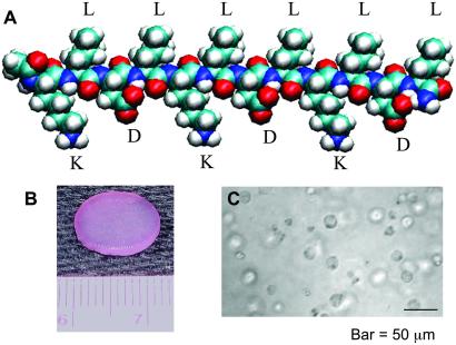
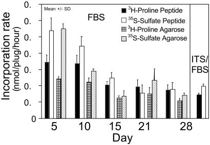
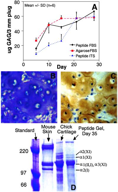
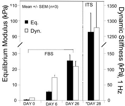
Emerging medical technologies for effective and lasting repair of articular cartilage include delivery of cells or cell-seeded scaffolds to a defect site to initiate de novo tissue regeneration. Biocompatible scaffolds assist in providing a template for cell distribution and extracellular matrix (ECM) accumulation in a three-dimensional geometry. A major challenge in choosing an appropriate scaffold for cartilage repair is the identification of a material that can simultaneously stimulate high rates of cell division and high rates of cell synthesis of phenotypically specific ECM macromolecules until repair evolves into steady-state tissue maintenance. We have devised a self-assembling peptide hydrogel scaffold for cartilage repair and developed a method to encapsulate chondrocytes within the peptide hydrogel. During 4 weeks of culture in vitro, chondrocytes seeded within the peptide hydrogel retained their morphology and developed a cartilage-like ECM rich in proteoglycans and type II collagen, indicative of a stable chondrocyte phenotype. Time-dependent accumulation of this ECM was paralleled by increases in material stiffness, indicative of deposition of mechanically functional neo-tissue. Taken together, these results demonstrate the potential of a self-assembling peptide hydrogel as a scaffold for the synthesis and accumulation of a true cartilage-like ECM within a three-dimensional cell culture for cartilage tissue repair.
Figures
References
-
- Brittberg M, Lindahl A, Nilsson A, Ohlsson C, Isaksson O, Peterson L. N Engl J Med. 1994;331:889–895. - PubMed
-
- Glowacki J. J Rehabil Res Dev. 2000;37:171–177. - PubMed
-
- Schuman L, Buma P, Versleyen D, de Man B, van der Kraan P M, van den Berg W B, Homminga G N. Biomaterials. 1995;16:809–814. - PubMed
-
- Nehrer S, Breinan H A, Ramappa A, Shortkroff S, Young G, Minas T, Sledge C B, Yannas I V, Spector M. J Biomed Mater Res. 1997;38:95–104. - PubMed
-
- Grande D A, Halberstadt C, Naughton G, Schwartz R, Manji R. J Biomed Mater Res. 1997;34:211–220. - PubMed
Publication types
MeSH terms
Substances
Grants and funding
LinkOut - more resources
Full Text Sources
Other Literature Sources
